Lupine Publishers | Journal of Otolaryngology
Abstract
Objective: To determine the correlation between clinical diagnosis (Squamous cell carcinoma or Lymphoma) and immunohistochemical results (Cytokeratin or Leukocyte Common Antigen) among patients with undifferentiated nasopharyngeal malignancy.
Study Design: Analytical, cohort study
Setting: Tertiary private hospital in Metro Manila Sample population and Methodology: Patients diagnosed of undifferentiated nasopharyngeal malignancy for the recent five (5) years were included in the study. Clinical diagnosis of nasopharyngeal squamous cell carcinoma or lymphoma was correlated with positive immunohistochemistry of cytokeratin (CK) and Leukocyte Common Antigen (LCA) respectively.
Results and Conclusion: The results showed that there was a significant association between the clinical diagnosis and immunochemistry results (p value 0.05). Among the 23 patients with clinical diagnosis of squamous cell carcinoma, only 19 patients (82.6%) were confirmed with CK reactivity while 4 cases were reactive with LCA. Among the 7 patients with clinical diagnosis of Lymphoma, only 4 patients (57.1%) were confirmed with LCA reactivity while 3 were positive for cytokeratin. A total of 22 patients were interpreted as cytokeratin positive (73.7%) and 8 patients were LCA positive (26.3%). Hence, there was a relatively fair association between clinical diagnosis of lymphoma and confirmatory LCA immunostain.
Keywords: Nasopharyngeal cancer; undifferentiated carcinoma; squamous cell cancer; lymphoma; LCA, cytokeratin
Abbreviations:LCA: Leukocyte Common Antigen; CK: Cytokeratin; IHC: Immunohistochemistry; NPCA: Nasopharyngeal Carcinoma
Introduction
Diagnosis of nasopharyngeal cancer depends upon a good history, physical examination findings and ancillary procedures. Nasopharyngeal lesions whether squamous cell carcinoma or lymphoma may manifest with epistaxis, middle ear effusion and neck nodes early in the disease process while cranial nerve deficits such as abducens nerve palsy or diplopia are late manifestations [1]. It has to be emphasized that in order to arrive to an accurate diagnosis, a good history taking is indispensable as well as a discerning clinical eye. This is essential because a correct diagnosis will determine the appropriate course of treatment regimen of a particular case. Nasal endoscopy is usually done to assess and ascertain any abnormalities in the nasal cavity and nasopharyngeal vault. Tissue obtained from nasopharyngeal punch biopsy is done to confirm the diagnosis. When histopathologic result would reveal undifferentiated carcinoma, this would require immunohistochemical analysis to determine whether it is squamous cell carcinoma or lymphoma. A local study by Cachuela in 2007, showed the value of Leukocyte Common Antigen (LCA) and Cytokeratin (CK) immunohistochemical stains in establishing a definitive diagnosis in undifferentiated tumors of the head and neck. It was a descriptive study and the correlation of clinical diagnosis and immunohistochemistry result was not determined [2]. Establishing the definitive diagnosis in undifferentiated tumors of the nasopharynx is crucial because the cell lineage of origin differs entirely as well as the modality of treatment. Hence, it would be wise and imperative to determine the correlation between the clinical diagnosis of SCCA and lymphoma and immunohistochemistry results (Cytokeratin and Leukocyte Common Antigen) respectively. This will help determine and evaluate the accuracy of the clinical eye in diagnosing patients with squamous cell carcinoma and lymphoma of the nasopharynx compared with the final immunohistochemistry results.
Review of Related Literature
Immunohistochemistry (IHC) is the process whereby
antibodies are used to detect antigens. Major components in a
complete immunohistochemistry are:
a) Primary antibody binds to specific antigen.
b) Antibody-antigen complex is formed by incubation with a
secondary, enzyme-conjugated, antibody.
c) With presence of substrate and chromogen, the enzyme
catalyzes to generate colored deposits at the sites of antibodyantigen
binding.
This can be done with simple staining, immunofluorescence, immunoenzymological staining or immunocolloidal gold technique [3]. Immunological methods can now resolve the majority of difficulties arising over the histological diagnosis of “unclassifiable” tumors. In immunohistochemistry, the features evaluated are location of immunoreactivity, identification of immunoreactive cells, and intensity of immunoreactivity [2]. The LCA has membranous tumor immunoreactivity while CK has cytoplasmic tumor immunoreactivity. Immunostaining using LCA and CK to confirm undifferentiated tumors in the head and neck allows definitive diagnosis of either carcinoma or lymphoma by 100% [2]. Validation of immunohistochemical stains for their specificity and sensitivity studied by Kutin [4], Michels [5] and Wick [6] showed that LCA is an excellent cell marker to distinguish hematopoietic neoplasms, particularly of the lymphoid type, from poorly differentiat tumors of epithelial, mesenchymal and neural derivation. The specificity of the LCA immunostaining technique was 100% and its sensitivity was 96%. In a study by Michie [7] to distinguish undifferentiated tumors with a histologic differential diagnosis of carcinoma versus lymphoma using immunostain anti-CK and anti- LCA, the results indicate specificity of 100% and sensitivity of 86%. Hence, correlating clinical diagnosis of squamous cell carcinoma and lymphoma with CK and LCA respectively would be necessary to determine the accuracy of clinical impression.
General objective
To determine the correlation between clinical diagnosis (Squamous cell carcinoma or lymphoma) and immunohistochemical results (Cytokeratin or Leukocyte Common Antigen) among patients with undifferentiated nasopharyngeal malignancy in a tertiary hospital in Metro Manila.
Specific objectives
a) To determine the demographic distribution of the
population according to age and sex distribution, chief
complaint, signs and symptoms and ENT findings.
b) To determine the incidence of squamous cell carcinoma
and lymphoma among undifferentiated nasopharyngeal
tumors.
c) To determine the immunologic reactivity to leukocyte
common antigen (LCA) and cytokeratin (CK) immunostain in
patients diagnosed with undifferentiated carcinomas of the
nasopharynx.
Sample Population & Methodology
Research Design
This is an misperceive, cohort study with prospective and retrospective enrolment. The sample population included review of old and new cases of patients with undifferentiated carcinoma of the nasopharynx.
Sample Population
Patients who consulted or were admitted at the MCU-FDTMF Hospital under the department of ENT Head and Neck Surgery for the past five (5) years, with a histopathologic diagnosis of undifferentiated carcinoma of the nasopharynx were included in this study. The consultants were informed about the study and patients were referred to the investigator accordingly.
Exclusion Criteria
a) Patients with metastatic nasopharyngeal mass from other
primary undifferentiated carcinoma.
b) Absence of immunohistochemistry results (LCA and CK)
for those patients seen / admitted in the past.
The patients’ clinical history with emphasis on ENT complaints such as nasal obstruction and / or epistaxis, aural fullness and effusion, smoking history were recorded. If nasopharyngeal mass was seen on endoscopy, punch biopsy was done, and the specimen was sent for histopathology. If the result was undifferentiated carcinoma, the patient would be included in the study and the specimen was sent for immunohistochemistry (CK and LCA) for confirmation of the diagnosis. The patient was informed about the study and informed consent was secured.
Sample Size
Using the 95% confidence level, the ideal sample size was computed to be n=73. However, due to the limited number of cases, the sample size is smaller and would only be limited to the number of cases seen during the period covered. It has to be stated that this is a limitation of this study.
(Zα)2pq
where Zα =1.96 at 95% confidence interval 95% = 0.95
N = ---------------
p = accuracy
E2q = 1 – p
1-0.95 = 0.05
E = error of 5% 0.05
Results
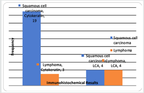
Out of 84 patients with endoscopic findings of nasopharyngeal mass who underwent punch biopsy, only 30 with histopathologic result of undifferentiated carcinoma were included in the study. Others were clinically diagnosed with adenoiditis (16.7%), tuberculosis (18.5%), salivary gland malignancy (3.7%), nasopharyngeal carcinoma (44.4%) and lymphoma (16.7%). The age range is 19 to 65 years old and composed of 26 male (86.7%) and 4 females (13.3%). Qualitative variables, which include the clinical diagnosis and immunostaining results, were tabulated in a contingency table and analyzed using Chi-square test to determine if the clinical diagnosis is associated with the immunohistochemical result. The results showed that there was a significant association between the clinical diagnosis and immunochemistry results (p value of 0.05) (Table 1). Significantly, more proportion of subjects with squamous cell carcinoma are cytokeratin positive and those with lymphoma are LCA positive. Among the 23 patients with clinical diagnosis of squamous cell carcinoma, only 19 patients (82.6%) were confirmed with CK reactivity while 4 cases were reactive with LCA. Among the 7 patients with clinical diagnosis of Lymphoma, only 4 patients (57.1%) were confirmed with LCA reactivity while 3 were positive for cytokeratin. Hence, a total of 22 patients were interpreted as cytokeratin positive (73.7%) and 8 patients were LCA positive (26.3%) (Table 2) (Figure 1).
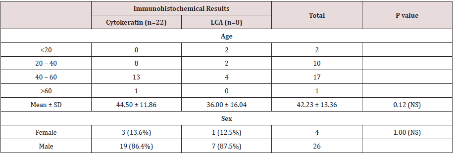
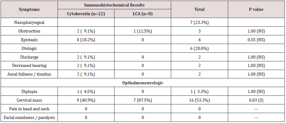
Table 3 shows the association of age and sex with immunohistochemical results. Majority of the patients with positive cytokeratin and LCA results belong to the 40-60 age group. Two patients less than 20 yrs old were positive for LCA. Men are affected more frequently than women in both squamous cell carcinoma (19:3) and lymphoma (7:1). The most common clinical manifestation of nasopharyngeal squamous cell carcinoma includes presence of cervical mass (40.9%), followed by nasopharyngeal complaints such as nasal obstruction and epistaxis (27.3%), and otologic symptoms (27.3%) such as decreased hearing, aural discharge, and aural fullness. One patient presented with diplopia (4.5%). The majority of patients with lymphoma presented with bilateral neck nodes (87.5%), with one complaining of chronic nasal obstruction (12.5%). Table 4 shows the association of clinical signs/ symptoms with immunohistochemical results. The results showed that there was no significant association (p values >0.05) except for cervical mass (p=0.03) which was present among patients who were LCA positive than those with Cytokeratin (Table 5).
Discussion
Nasopharyngeal carcinoma (NPCA) is common in many oriental
countries in any age group and is a relatively rare neoplasm among
Caucasians. The predilection of NPCA among Asian countries may
be attributed to the dietary regimen which is made up of salty fish
and fermented foods which is high in volatile nitrosamines [1].
There are different types of malignancy which may develop in the
nasopharynx which include squamous cell carcinoma, lymphoma,
salivary gland malignancy, and sarcomas [8]. The most common are
squamous cell carcinomas, which can be divided into 3 types of cell
differentiation (well, moderately or poorly differentiated), and are
collectively called nasopharyngeal carcinoma. The nasopharynx is
lined by either stratified squamous epithelium or pseudostratified
columnar epithelium where squamous cell carcinoma commonly
originates. The nasopharyngeal mucosa also contains salivary and
lymphoid tissues, so that salivary tumor and lymphoma may develop
although less frequently [9]. The gross appearance of the mass is
usually descriptive of the type of malignancy that is involved; a
smooth bulging nasopharyngeal mass is noted in lymphoma while a
friable corrugated appearance of the mass, infiltrative or exophytic,
is commonly seen in squamous cell carcinoma [1,9]. Among the
patients with squamous cell carcinoma, 8 presented with bulging
friable corrugated mass, 7 with exophytic smooth lobulated mass,
4 with ulcerative friable mass, 3 with infiltrative friable slightly
elevated mass. All patients with lymphoma presented with smooth
bulging lobulated nasopharyngeal mass.
Due to the hidden anatomic location, nasopharyngeal carcinoma
may remain clinically silent. This will explain why nasopharyngeal
lesions are considered areas of silent primaries. It arises more
frequently in the superior portion, followed by pharyngeal recess
(fossa of Rosen Mueller) in the lateral wall. Clinical manifestations
include cervical masses 77%; nasopharyngeal problems consisting
of obstruction and epistaxis 64%; otologic complaints ranging
from serous discharge, otalgia, tinnitus and decreased hearing
secondary to Eustachian tube dysfunction 60% [10,11]. With
disease progression, cranial nerve involvement becomes apparent
such as ophthalmoneurologic manifestations presenting as
blurring of vision, diplopia and ptosis 34%.10,11 In this study, the
most common manifestation of nasopharyngeal squamous cell
carcinoma include presence of cervical mass (40.9%), followed by
nasopharyngeal complaints such as nasal obstruction and epistaxis
(27.3%), and otologic symptoms (27.3%) such as decreased hearing,
aural discharge and aural fullness. One patient presented with
diplopia (4.5%). The World Health Organization (WHO) histologic
system is used for the nomenclature of nasopharyngeal carcinoma,
which encompasses nonkeratinizing carcinoma (differentiated
or undifferentiated), keratinizing squamous cell carcinoma and
basaloid squamous cell carcinoma [1].
Undifferentiated carcinoma (previously WHO III) is composed
of cells with indistinct margins and round to oval nuclei with
prominent, round nucleoli. The cells tend to grow in a syncytium
rather than having a stratified or pavemented appearance. The
tumor can grow in well-defined epithelial aggregates (Regaud
pattern) descriptive of a carcinoma. It grows as ill-defined sheets,
small clusters, or individual cells admixed with lymphocytes
(Schmincke pattern). It is most often found in children, has a strong
correlation with EBV, tends to disseminate, and has a good response
to irradiation [12]. In this study, out of the 30 patients diagnosed
of undifferentiated carcinoma, 22 (73.3%) were interpreted as
cytokeratin positive. The clinical signs of Lymphoma in the head
and neck are frequently diverse. The sites of involvement can be
subdivided into (1) nodal; (2) extra nodal, lymphatic; and (3) extra
nodal, extra lymphatic. Extra nodal, lymphatic lymphoma refers to
tumor found in Waldeyer’s ring. The extra nodal, extra lymphatic
category includes all NHL not in nodes or Waldeyer’s ring, with
the areas of involvement including the orbit, sinus, nose, mandible,
deep facial spaces, parotid gland, and dermis. Nodal lymphoma
is the most common. When nodes are located in sites atypical of
the usual drainage routes for squamous cell carcinoma such as
the submental, submandibular, jugular chain, retropharyngeal, or
posterior triangle nodal chains, lymphoma should be considered.12
Of the 30 patients with histopathologic diagnosis of undifferentiated
carcinoma, 8 (26.7%) were interpreted as LCA positive. In our
study, the majority of patients with lymphoma presented with neck
nodes (87.5%), with one complaining of chronic nasal obstruction
(12.5%). Most presented with bilateral neck nodes (75%), mostly
in the jugular chain. Only one patient presented with submental
and submandibular nodes (12.5%). The majority belong to
the 40-60 age group, with two patients less than 20 yrs old. In
most instances, the differentiation of a tumor can be classified
morphologically with certainty by histological examination. When
the dilemma of undifferentiated carcinoma or unclassifiable tumor
arises, immunohistochemical staining helps resolve this issue.
The introduction of hybridoma technology and the development
of monoclonal antibodies directed against cellular components
have generated improved methods of identifying undifferentiated
tumors.
The goal of such technique is the development of an antibody
directed against a specific cell or cellular component such as
cytkeratin (CK) and epithelial membrane antigen (EMA) for
squamous cell carcinoma; S-100, keratin, vimentin, desmin and CEA
for salivary glands; and leukocyte common antigen (LCA) and OKT
(CD) antigens for lymphoma. Among the 23 patients with clinical
diagnosis of squamous cell carcinoma, only 19 patients (82.6%)
were confirmed with CK reactivity while 4 cases were reactive with
LCA. Among the 7 patients with clinical diagnosis of Lymphoma,
only 4 patients (57.1%) were confirmed with LCA reactivity while 3 were
positive for cytokeratin. Hence, a total of 22 patients were
interpreted as cytokeratin positive (73.7%) and 8 patients were LCA
positive (26.3%). There were patients who turned out positive for
cytokeratin who presented with multiple neck nodes with smooth
lobulated or only slightly elevated nasopharyngeal mass, hence, the
initial impression was lymphoma. Patients with clinical impression
of squamous cell carcinoma, on the other hand, who were reactive
to LCA, had history of chronic nasal obstruction or decreased
hearing / aural fullness, which are frequently seen in patients
with squamous cell carcinoma. Both LCA and CK immunostain are
specific (100%) and sensitive (86-95%) to lymphoid and epithelialderived
tissues respectively [2]. Squamous cell carcinoma in general
is responsive to radiation therapy in contrast to lymphoma which is
treated with chemotherapy.8 Arriving at the appropriate histologic
diagnosis is important to guide medical practitioners in choosing
the appropriate treatment.
Conclusion
The results showed that there was a significant association between the clinical diagnosis and immunochemistry results (p value 0.05). Among the 7 patients with clinical diagnosis of Lymphoma, only 4 patients (57.1%) were confirmed with LCA reactivity while 3 were positive for cytokeratin. A total of 22 patients were interpreted as cytokeratin positive (73.7%) and 8 patients were LCA positive (26.3%). There was a relatively fair association between clinical diagnosis of lymphoma and confirmatory LCA immunostain. A good clinical history and physical examination still has an integral part in the diagnosis of nasopharyngeal carcinoma. Immunostaining is necessary to confirm the diagnosis.
Read More Lupine Publishers Otolaryngology
Journal
Articles:
https://lupine-publishers-otolaryngology.blogspot.com/


No comments:
Post a Comment