Friday, January 14, 2022

Lupine Publishers | Hearing Problems in Children and Mowat-Wilson Syndrome: A Case Report

 Lupine Publishers | Journal of Otolaryngology


Keywords: Deafness; dysmorphia; epilepsy; molecular study; MowatWilson syndrome

Abbreviations:MSW:Mowat-Wilson syndrome; AEP: Hearing Potentials

Introduction

Hearing problems, more precisely hearing loss in children, is a very frequent reason for consultation these days. In early childhood, these disorders affect language and communication, and deafness is the most common sensory deficit. According to the WHO, the hearing-impaired child is one whose hearing acuity is insufficient to enable him to learn his own language, to participate in the normal activities of his age and to follow with profit general school education [1]. Its prevalence is estimated between 1 and 3.4 per thousand at birth [2] and could reach 1 per 100 in cases of severe perinatal disease and the genetic origin is then implicated in 60-80% of cases [3]. But the etiologies differ, and hypoacusis can even be a symptom of congenital syndromic disease.

Case Study

Figure 1: Hypertelorism; large eyebrows; prominent columella.

Lupinepublishers-openaccess-otolaryngology-journal

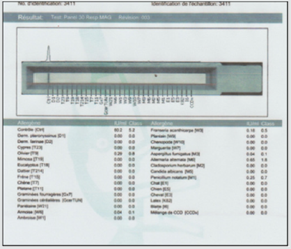
In our work we report the case of a 3-year-old girl from Algeria brought by her parents to Tunisia to take charge of hearing loss, according to them; presenting a language delay that has progressed for 2 years; with a particular dysmorphic facies and unexplored convulsive seizures. The interrogation clarified an outconsanguineous marriage; normal course of pregnancy with an uneventful vaginal delivery (according to the health record) with a birth weight of 3,400 kg. moreover, the child’s vaccination schedule was respected. The history of the disease dates back to the age of 18 months when the parents noticed delayed speech as well as hearing loss on call with the onset of seizures. , The clinical examination beginning with the inspection showed marked hypertelorism especially in the upper lip; high forehead with two frontal bumps; large eyebrows poor in their middle, saddle nose and prominent columella (Figure 1); large raised lobes with a central depression (Figure 2); blue staining of the eardrums on otoscopy with congestive appearance of the nasal mucosa and hypertrophy of the inferior turbinate’s. Nasofibroscopy performed eliminated the presence of adenoids. The rest of the examination is normal with a weight of 18.4 kg and a blood pressure of 100/60 mmHg. Additional audiometric examinations have revealed bilateral serummucous otitis with a flat line on the tympanometry and a normal threshold at PEA and deafness transmission of 30 DB to behavioral audiometry. A specific assay of specific pediatric pneumallergenic IgE revealed a large number of allergens in children (Figure 3). A medical treatment based on oral corticosteroid therapy at a dose of 2 mg / kg associated with antibiotic therapy (clavulanic acid12.5 mg + amoxicill100 mg) at a dose of 80 mg / kg and local nasal treatment based on corticosteroid therapy and nasal washings with sprays based on essential oils. In the meantime, we have further explored within the framework of the exploration of epilepsy and the particular dysmorphic facies.

Figure 2: Large earlobes.

Lupinepublishers-openaccess-otolaryngology-journal

Figure 3: Dosage of specific pneum allergenic IgE positive.

Lupinepublishers-openaccess-otolaryngology-journal

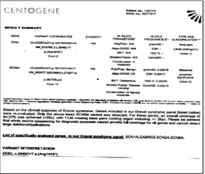
An EEG performed showed a diffuse slowing of the background rhythm with brief generalized spikes. MRI of the brain and ponto-cerebellar angles returned strictly normal especially no microcephaly or agenesis of the corpus callosum and an intact inner ear. Before epilepsy and dysmorphic facies, a congenital syndrome was suspected and the first that came to mind was Dravet’s syndrome, given the generalized epileptic seizures, hence a genetic study (total sequencing of the exome) of the child was performed revealing the diagnosis of Mowat-Wilson syndrome (Figure 4). A second genetic study of the parents’ genome was made and returned without suspicious abnormalities. The now multidisciplinary care (neuropediatrician, speech therapist and occupational therapist) has led to good further development; sedation of convulsive seizures under an anti-epileptic drug at a dose of 500 mg / day and language acquisition around the age of 3 years 6 months and resumption of an active life with the children (patient put in daycare; follows swimming at the pool).

Figure 4: Sequencing of the exome.

Lupinepublishers-openaccess-otolaryngology-journal

Discussion

Childhood deafness is a very common health problem, usually associated with delayed or impaired speech. All origins combined; deafness affects 1.3 to 2.3 children out of 1000 in industrialized countries. Deafness can be classified into 2 categories according to the mechanism [2,4]. Conductive hearing loss linked to damage to the outer ear or the middle ear. They are acquired in 99% of cases and are most often accessible to medical treatment +/- surgical. Their etiology is dominated in young children by inflammatory and infectious pathologies linked to tubal dysfunction as it is in our case. They can also be secondary to malformative, traumatic or tumor pathologies of the pinna, the external auditory canal or the ossicles. This deafnessis most common in children and do not exceed 60dB of hearing loss; 95% of this deafness are linked to otitis media. Congenital conductive hearing loss accounts for 0.5% of conductive hearing loss.
Major aplasia: The frequency of debilitating bilateral forms varies greatly depending on the series, probably in the order of 20 to 30%. Four grades have been described from turbinate ear (grade I) to anotia (grade IV) resulting in conductive hearing loss of 60 to 70 dB. Minor aplasia: the malformation of the middle ear is isolated or associated with minor malformations of the outer ear or certain polymalformative syndromes (in our case large lobe with a well calibrated external auditory canal).

Sensorineural hearing loss which may be secondary to pathology of the organ of Corti, the auditory nerve and / or central auditory areas [5]. Hearing loss is extensive from mild to complete and is very commonly associated with message distortions. This deafnessis in two thirds of the cases genetic, autosomal recessive in 80% of the cases, autosomal dominant in 20% of the cases, linked to the X affecting only rare boys, or mitochondrial (deaf mother transmitting to all her children ) exceptional. The consequences of this high prevalence make it possible to carry out a routine molecular diagnosis and to confirm the genetic nature of deafness, thus giving the parents the risk of recurrence (25%) for future births. Whatever the mode of transmission, sensorineural hearing loss is either isolated (two thirds of cases) or associated with a polymalformative syndrome. Radiological exploration (MRI) associated with a study of hearing potentials (AEP) associated with a genetic molecular study confirms the diagnosis. In our case, the cochlear and genetic origin was eliminated (normal inner ear and PEA). Mixed deafness which results from damage to the outer and / or middle and inner ear.

Deafness can be classified according to the date of onset [4,6]
a) The audio-phonatory loop is necessary for the development of the child’s language and speech, its interruption therefore does not have the same consequences depending on the age of onset of deafness.
b) Congenital deafness: deafness exists from birth.
c) Pre-lingual deafness (where our case is; hearing loss since the age of 18 months): occurring before the age of 2 years, are the most serious because the early acquisitions essential for subsequent language development do not take place.
d) Perilingual deafness:Occurring between 2 and 4 years e) Postlingual deafness:Itoccurs after the age of 4 years.

The careful etiological assessment is therefore an essential step for many reasons: genetic counseling only becomes possible with a precise diagnosis, especially in the face of deafness associated with a polymalformative syndrome and the progressive prognosis and the management will subsequently depend on the cause ; as in our case we followed the research protocol for a congenital syndromic etiology which led us, to everyone’s surprise, to a rare syndrome still under study: Mowat-Wilson syndrome (MWS) which is a syndrome Multiple birth defects characterized by a distinct facial phenotype, intellectual disability, epilepsy, Hirschsprung’s disease (HSC), and variable birth defects. The prevalence is estimated between 1 / 50,000 and 1 / 70,000 live births [7]. More than 200 patients have been reported to date. It seems likely that MWS is underdiagnosed, especially in patients without HSC.The typical characteristic facies of MWS includes a high forehead, frontal boss, large eyebrows flared medially and sparse in the middle part, hypertelorism, deep but large eyes, wide and raised earlobes with a central depression, a saddle nose with a prominent rounded nasal end, prominent columella, open mouth with M-shaped upper lip and prominent but narrow and triangular pointed chin (same description for our patient). The facies become more pronounced with age. The associated HSCR causes constipation which frequently persists after surgery. Patients generally have moderate to severe intellectual disability.

Speech is absent or limited to a few words, with an onset around 5-6 years old. Seizures are common; all types of seizures (absence, generalized tonic-clonic seizures, myoclonic and focal seizures) have been reported. Most of the subjects have a cheerful demeanor with frequent smiles and a sociable personality. Other associated congenital anomalies include cerebral anomalies (agenesis of the corpus callosum), heart (persistence of the ductus arteriosus, interventricular communication,pulmonary stenosis, valve), pulmonary (arterial sling, with or without tracheal stenosis), genitourinary (hypospadias, cryptorchidism, vesicoureteral reflux and hydronephrosis), ocular (microphthalmia) [in our case high myopia] and musculoskeletal (flatfoot, calcaneovalgus) This syndrome is caused by heterozygous mutations or deletions in the zinc finger E box binding gene for homeobox 2, ZEB2, (2q22.3) previously called ZFHX1B (SIP1) [8,9]. To date, over 100 deletions/ mutations have been reported in patients with a typical phenotype; these are often whole gene deletions or truncated mutations, suggesting that haploinsufficiency is the main pathological mechanism. Genotype-phenotype analysis studies show that facial gestalt and retarded psychomotor development are constant clinical features, while frequent and severe birth defects are variable. In a small number of patients, unusual mutations can lead to an atypical phenotype. The clinical diagnosis is then based by summarizing according to Zweier et al. [10] on the distinct facies and the ZEB2 mutational analysis.

Congenital malformations and seizures require early clinical investigation with the intervention of several specialists (including neonatologists and pediatricians) if the diagnosis is suspected early. In addition, prenatal diagnosis has become available and essential for subsequent pregnancies of parents with an affected child.The majority of cases of MWS reported so far were sporadic as in our case, however, germline mosaicism has been described. and the risk of recurrence was calculated at 2% [11] (his younger brother is strictly normal) Psychomotor development is delayed in all patients (ditto for our case), which is why rehabilitation (physiotherapy, psychomotricity and speech therapy ) should be started as soon as possible (for us, we started it as soon as the diagnosis made at the age of 3 years 2 months. Mortality and morbidity depend on elsewhere about the presence and severity of birth defects. It has been reported according to the literature that patients live in early adulthood but need help with activities of daily living.

Conclusion

Hearing is an infinitely precious sense for a child. It allows not only to perceive the outside world, but also to access information and communication. Any impairment of hearing function constitutes a handicap with significant socio-professional repercussions.Any suspicion of hypoacusis in the child must lead to an otolaryngological (ENT) examination and audiometric examinations appropriate to his age to rule out or confirm the hearing loss and, in the latter case, specify the mechanism and the degree and further exploration if there are any associated dysmorphic signs while keeping in mind the variability of congenital syndromes as it was in our case; knowing that Mowat-Wilson syndrome (SMW) is a complex congenital syndrome Described in 1998 by Mowat et al. with a prevalence still unknown and under study.

Read More Lupine Publishers Otolaryngology Journal Articles:
https://lupine-publishers-otolaryngology.blogspot.com/


Saturday, January 8, 2022

Lupine Publishers | Study of Brainstem Evoked Response Audiometry to Assess Hearing Loss in High Risk Children Below 5 Years

 Lupine Publishers | Journal of Otolaryngology


Abstract

Early detection of hearing loss and intervention is very important for the development of normal speech and language. A prospective study of brainstem evoked response audiometry to access hearing loss in high risk children below 5 years of age, to find out the incidence of hearing loss in children from high risk category and to determine the threshold of hearing by brainstem evoked response audiometry in high risk children by observing wave ‘V’ at the minimum intensity of click stimulus. 100 children having one or more high-risk factors for hearing loss (up to 5 years of age) according to the inclusion criteria had been selected for the study from the outpatient department. This study showed that BERA was found to be a useful, accurate tool to detect the type of hearing loss and the site of defect in auditory transmission of sound. This aids in planning the mode of intervention for early rehabilitation for speech and language development.

Keywords: Brainstem evoked response audiometry; hearing loss; high risk children

Introduction

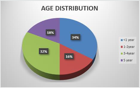
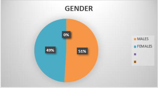
Early detection of hearing loss and intervention is very important for the development of normal speech and language. In India, 63 million people (6.3%) suffer from significant auditory loss. Four in every 1000 children suffer from severe to profound hearing loss. With over 100,000 babies that are born with hearing deficiency every year approximately 50% of all cases of congenital hearing loss are attributable to environmental factors such as congenital hyperbilirubinemia, ototoxic medication exposure, neonatal hypoxia, viral infections, and meningitis. The other 50% of cases are thought to be inherited, i.e., of genetic causes. Of these hereditary cases approximately 30% are classified as syndromic [1]. In a developing country like India newborn hearing screening is yet to be implemented. Joint committee on infant hearing recommends BERA (brainstem evoked response audiometry) technology as the only appropriate screening technique for use in the NICU (neonatal intensive care unit) (Figures 1-3). For infants who do not pass BERA testing in the NICU referral should be made directly to an audiologist for rescreening and when indicated comprehensive evaluation including diagnostic BERA should be done [2]. BERA is a far-field recording of the synchronized response of numerous neurons in the auditory pathway within the brainstem. BERA was first described by Sohmer and Feinmesser in 1967. It reflects neural activity generated at the level of the brainstem because of which it is not strongly affected by attention, sleep, or sedation [3].

Figure 1: Showing age distribution.

Lupinepublishers-openaccess-otolaryngology-journal

Figure 2: Showing sex distribution.

Lupinepublishers-openaccess-otolaryngology-journal

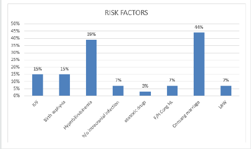
Figure 3: Showing risk factors among subjects.

Lupinepublishers-openaccess-otolaryngology-journal

Objectives

a) To find out the incidence of hearing loss in children from high risk category by brainstem evoked response audiometry.
b) To determine the threshold of hearing by brainstem evoked response audiometry in high risk children by observing wave ‘V’ at the minimum intensity of click stimulus.

Materials and Methods

100 children having one or more high-risk factors for hearing loss (up to 5 years of age) according to the inclusion criteria had been selected for the study from the outpatient department of Bangalore Medical College and Research Institute, Bangalore for from August 2019 to July 2020. General physical examination was conducted to know the health status and to rule out any deformities in the subjects. Any local pathology present in ear, nose and throat was treated and BERA was carried out in all children.

Inclusion criteria

Following high risk children are included in the study
a) History of intrauterine infections (congenital infections) b) Birth weight < 1.5 kgs.
c) Hyperbilirubinemia > 20mg/dl.
d) Moderate to severe birth asphyxia.
e) Maternal or neonatal history of ototoxic drug intake.
f) Documentary evidence of intracranial infections in the past.
g) Family history of childhood hearing impairment.
h) Consanguineous marriage.

Exclusion criteria

a) Known case of seizure disorders where BERA cannot be interpreted
b) Atresia or stenosis of external ear canal of both ears.
c) Patients not giving consent for BERA.

Parameter studied

BERA threshold for each ear with absolute latencies of wave I, II, III, IV and V waves interpeak latencies (IPL) of I-III, I-V and III-V and amplitude ratio V/I were considered from the recordings of high risk children.

Procedure

It was performed in dust free, soundproof, air-conditioned room free from electromagnetic disturbances. Syrup Pedichlor (25 mg/kg body wt.) was given to sedate the baby half an hour before procedure. After cleaning forehead, vertex and both mastoid regions with Nuprep (skin prep gel), surface electrodes were placed at the vertex, both mastoids and forehead (ground) and subject was tested in sleeping state with neck slightly flexed to minimize any myogenic activity. Headphone (TDH-39) was held against the ear of baby taking care that external auditory canal does not collapse. 2000/4000 clicks at a rate of 11.1/sec was given and the graphs was recorded on BERA machine Labat Epic- Plus.

Results

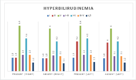
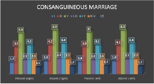
In this study 34% were in the age group <1 year. 51% were males and 49% were females. Almost equal number of males and females. In the study 15% had intrauterine infections, out of 15 subjects, 7 of them had CMV and 8 of them had Rubella. 39% had Hyperbilirubinemia, 44% had consanguineous marriage, of them 18 had 2nd degree marriage and 26 had 3rd degree marriage. There was significant difference in mean BERA parameters (V(dB) Threshold, III, V, I-III, I-V, III-V between those with and without hyperbilirubinemia. The above said parameters were significantly high among those with hyperbilirubinemia in right and left ear. There was Significant difference in mean V(dB) Threshold, III, V and I-V between those with and without H/o Consanguineous Marriage in Right ear. There was significant difference in mean V (dB) Threshold, V b/w those with and without H/o Consanguineous Marriage in Left ear. Those with Consanguineous Marriage, majority had mild (35.7%) hearing loss (Figures 4 & 5). In this study there was statistically significant difference between wave V threshold and high-risk factors consanguineous marriage and hyperbilirubinemia. There was no significant difference in mean BERA parameters between those with and without intrauterine infections, low birth weight, intrauterine infections, h/o intracranial infections, h/o ototoxic drug intake, family h/o congenital hearing loss and birth asphyxia in both right and left ear. 26 out 44 children with history of consanguineous marriage had hearing loss out of which 4 were unilateral with mild hearing loss. 34 out of 39 children with hyperbilirubinemia as a risk factor had hearing loss with one child having unilateral hearing loss.

Figure 4: Bar diagram showing Comparison of BERA parameters with respect to presence and absence of hyperbilirubinemia in Right and Left ear.

Lupinepublishers-openaccess-otolaryngology-journal

Figure 5: Bar diagram showing Comparison of BERA parameters with respect to presence and absence of consanguineous Marriage in Right and Left ear.

Lupinepublishers-openaccess-otolaryngology-journal

Discussion

Early detection of hearing loss and intervention is very important for the development of normal speech and language. Majority of children with hearing loss belong to age group 1-5 years between which most of the speech and language development occurs.4 Interventions usually vary from sign language to cochlear implantation. Brainstem evoked response audiometry although more time-consuming, is an accurate and reliable test for early detection of neural conduction abnormalities in the auditory pathway from the cochlea till the brainstem. The reported sensitivity of the BERA for hearing assessment was 100% and specificity around 86% [5]. Earlier perinatal causes like birth asphyxia, hyperbilirubinemia, and intrauterine infections were on the rise but recently inherited causes are on the rise especially in developing countries like India. This is probably due to better treatment facilities and referral centers available. In our study the most common risk factor is shown to be consanguineous marriage showing inherited causes were on the rise. Abnormality at the level of cochlear nucleus and brainstem due to inheritance of abnormal genes. This data is consistent with studies by Ramanathan [4] and Mallikarjun [6] where inherited causes were on the rise. Health education is of prime importance to increase awareness of the detrimental health effects of consanguineous marriage.
The next most common risk factor was found to be hyperbilirubinemia having Wave V absolute latency was increased in both the ears and also interpeak latency I-V [7]. This indicates that raised bilirubin levels is a important risk factor for detecting hearing loss.8 In most infants with raised bilirubin levels the values reverted back to normal after treatment indicating that it is a reversible cause and prompt treatment is required. This is similar to a study by Pramod Sharma [7] and by other authors [8-10] in which there was statistically significant correlation in prolongation of latency and the interpeak latencies with serum bilirubin levels more than 25 mg%. Follow up of infants with raised bilirubin levels having hearing loss is required. Children with birth asphyxia could have hearing loss due to the depressed end cochlear potential induced by hypoxia and acidosis causing the involvement of cochlea or cochlear nerve showing dysfunction in the peripheral auditory process [11,12]. A definitive conclusion cannot be made out regarding the outcome of the study, but it does suggest that all high-risk children will benefit from early detection of hearing loss and rehabilitation through BERA. High risk children should be screened in hospital NICUs, prior to discharge from the hospital and immunization clinics particularly around 6 months of age or below one year using BERA for timely intervention to avoid delay in speech and language development. A national program dedicated to detecting early hearing loss would help reducing the burden of handicap by avoiding delay in intellectual development of the child. Brainstem evoked response audiometry though expensive is a safe and accurate procedure to ascertain whether the hearing loss can benefit through common rehabilitative measures like hearing aids or not. Infants with abnormal BERA recordings should be rescreened within 3 months and several times within the first year. If abnormal responses persist immediate intervention to develop speech and language should be undertaken.

Conclusion

From our study we can thus conclude that among the high-risk factors, consanguineous marriage seems to be the most common factor causing hearing loss in children. This factor being is a preventable cause, as compared to others, by premarital genetic counseling and health education in lower socioeconomic groups. The next most common factor is shown to be hyperbilirubinemia. This is proven to be a reversible cause of hearing loss according to some studies. Therefore, early detection treatment and follow up is very important in such children. Almost all of the children with a family history of hearing loss had mild to moderate hearing loss suggesting inheritance of abnormal proteins through genes but the sample size for the same was less to derive any significant correlation. BERA was found to be a useful, accurate tool to detect the type of hearing loss and the site of defect in auditory transmission of sound. This aids in planning the mode of intervention for early rehabilitation for speech and language development.

Read More Lupine Publishers Otolaryngology Journal Articles:
https://lupine-publishers-otolaryngology.blogspot.com/

Choanal Atresia Repair, A Comparison Between Transnasal Puncture With Dilatation And Stentless Endoscopic Transnasal Drilling

  Abstract Background: in this study we present the outcome of surgical repair of choanal atresia of 33 patients underwent t...