Showing posts with label Journal of Otolaryngology research. Show all posts

Friday, October 16, 2020

Lupine Publishers | Lipoma of the Parotid Gland: A Case Report

 Lupine Publishers | Journal of Otolaryngology


Abstract

Lipoma of the parotid gland is extremely rare, accounting for only 0,6% - 4,4% of all parotid tumors. We present a rare case of lipoma of the superficial parotid lobe. A 68 - year-old man, presented in our department with a mass of the left parotid region. Clinical examination revealed a mobile, soft, non-tender mass in the area of the left parotid gland. MRI concluded to a lipoma of the superficial lobe of parotid gland. Parotidectomy preserving the facial nerve was performed. No complication nor recurrence were noted after a follow-up of 12 months. Lipoma arising in the parotid gland is extremely rare. Resection of this tumor requires full exposure of the facial nerve and its branches.

Keywords: Lipoma; Parotid gland; Superficial lobe; Parotidectomy

Introduction

Lipoma is a common mesenchymal soft tissue tumor that can be found in any part of the body. It can develop in the head and neck region in 15–20% of cases. Rarely it can arise in the Parotid gland with a ranging incidence from 0,6 to 4,4 % [1]. Clinical diagnosis may be difficult. MRI is necessary, in diagnosis [2]. Surgical management of these tumors is challenging and need meticulous dissection of the facial nerve. We report a case of lipoma arising of the parotid gland and we discuss through literature its clinical and therapeutic features.

Case Report

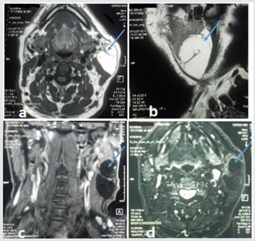
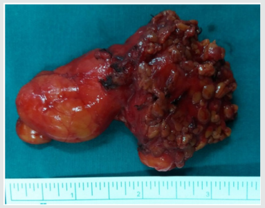
A 68 - year-old man presented with mass of the left parotid region, which was painless slow-growing for 5 years. Clinical examination revealed a mobile, soft, non-tender mass that measured about 6cm in diameter in the area of the left parotid gland, extending from the ear lobule to the left mandibular angle. There was no facial paralysis nor evidence of cervical lymphadenopathy. Magnetic resonance imaging (MRI) showed a well-defined homogeneous lesion of the superficial lobe of the left parotid gland with an enhanced signal on T1- and T2-weighted sequences and weak signal on fat suppressed sequences (Figure 1). The diagnosis of intra parotid lipoma was evoked. A left superficial parotidectomy preserving the facial nerve was performed. The specimen was soft, yellowish, well- circumscribed measuring 80*34mm. Histological examination revealed a well-circumscribed aggregate of mature adipocytes surrounded by a thin fibrous capsule confirming the diagnosis of intraparotid lipoma. No recurrence or complication were observed after a follow-up of 12 months.

Discussion

Lipoma is one of the most frequently encountered benign mesenchymal tumors that may originate from adipose tissue in any part of the body [3,4]. Rarely, it can develop in the parotid gland with reported incidence ranging from 0,6% to 4,4% among parotid tumors [3,5]. Lipoma may occur at any age, but most frequently between 40 and 60 years with a male predominance [5,6]. Its aetiology is unknown. It can be caused by heredity, obesity, diabetes, radiation, endocrine disorders, insulin injection, corticosteroid therapy and trauma [7]. In our case, we did not find any aetiological factors. Most of the reported cases were located at the superficial parotid lobe [7]. Lipomas involving the deep parotid lobe are extremely rare [5-8]. Clinical diagnosis may be difficult [9], especially for tumors located at the deep parotid lobe because it is difficult to evaluate the relationship between these masses and the surrounding tissues. Those situated at superficial parotid lobe usually appear as a slow growing, non-tender, movable and well-differentiated soft mass in parotid region [3-10]. Facial paralysis and pain are uncommon signs and rarely have been described [5-11]. This benign clinical presentation is most often mistaken for Warthin tumor or pleomorphic adenoma [3-12]. Fine needle aspiration cytology (FNAC) has great value in the diagnosis of parotid tumors and requires an experienced cytologist. Its accuracy drops to less than 50% in the cases of parotid lipomas [5-13]. On imaging, CT scan shows hypodense, homogeneous and well delineated mass with few septations and negative attenuation, without contrast enhancement [3-13]. However, CT scan cannot distinguish lipoma from surrounding adipose tissue. MRI remains the best diagnostic tool that can accurately diagnose lipomas [5-1]. Lipomas produce strong signals on T1- and T2 weighted sequences and weak signals on fat-suppressed sequences. After Gadolinium injection, the mass still hypointense to parotid, homogeneous in signal and uniformly non-enhancing [3]. MRI can also clearly define the limits of lipoma from normal adipose tissue and may be useful in determining the appropriate surgical approach.

Figure 1: MRI of the parotid gland showing homogeneous mass on the superficial lobe of the left parotid gland on hyper signal T1 and T2 and a weak signal on fat suppressed sequences. The lesion is hypointense to parotid and uniformly non-enhancing.

lupinepublishers-openaccess-journal-otolaryngology

Figure 2: Macroscopic findings of the resected tumor.

lupinepublishers-openaccess-journal-otolaryngology

Histopathologic investigation reveals mature adipose tissue separated from parotid gland parenchyma with a fibrous capsule. Identification of a capsule may aid in distinguishing such a neoplasm from pseudolipoma, lobular lipomatous atrophy, or lipomatosis, all of which are unencapsulated [3]. Surgery is the treatment of choice of parotid gland lipoma, but its modalities remain controversial [8-12]. It should be performed by experienced surgeons because of the need for meticulous dissection of the facial nerve branches. The postoperative esthetic and functional results should be the major concerns [1]. Some surgeons recommend simple enucleation of a superficial lobe parotid lipoma with a small border of healthy parotid gland parenchyma, as this is easy to perform because of the well-defined capsule. Other surgeons suggest that the surgical management of parotid lipoma should be the same as that for other parotid tumors [5]. However, it is well known that transient facial nerve dysfunction and Frey’s syndrome may occur as complications following surgical intervention for parotid tumors and should be explained to the patient before operation [5]. Facial nerve dysfunction ranged between 8.2 and 65% after parotid gland surgery for benign tumors [10]. Therefore, it requires efforts such as facial nerve monitoring to identify the facial nerve. Recurrence rate of parotid lipoma after adequate resection is very low. It has been reported in 5% in all cases when it is well-encapsulated [5-9]. Malignant transformation into liposarcomas has been reported in few cases in the literature [9]. Therefore, careful follow-up is recommended.

Conclusion

Lipoma of the parotid gland is a rare benign tumor, which should be considered in the differential diagnosis of parotid gland’s mass lesions. MRI is essential to locate the tumor, and to precise its relationship with the facial nerve. Their management is challenging. Definitive diagnosis can only be accomplished with histopathologic review.

Read More Lupine Publishers Otolaryngology Journal Articles:
https://lupine-publishers-otolaryngology.blogspot.com/
Read More Lupine Publishers blogger Articles:
https://lupinepublishers.blogspot.com


Saturday, October 26, 2019

Lupine Publishers | Occurance of Dizziness in patients with Tinnitus Complaint

Lupine Publishers | Journal of Otolaryngology

Abstract


Introduction: Dizziness is a regular complaint, often accompanied by other symptoms, especially tinnitus. Tinnitus is one of the three major otoneurological manifestations, alongside neurosensorial hearing loss and dizziness, being it, most of the times, the main complaint among patients. The relationship between the vestibular and cochlear system it’s rather known, many pathologies can originate from one of both systems.
Objective: Investigate the correlation between the symptoms of tinnitus and dizziness, analyzing the level of disturbance, the sensation of frequency (pitch) and intensity (loudness) of the tinnitus with dizziness complaint.
Methodology: A descriptive, observational and quantifying field study took place. 126 individuals with tinnitus complaint, from both sexes, were studied. The anamnesis was performed approaching audiological symptoms, the THI questionnaire was applied, as well as acuphenometry.
Results: 71 individuals (56,3%) referred to dizziness complaints associated with the tinnitus; women represented a larger number (41,3%) (p=0,017). In regards of the level of disturbance of the tinnitus, most of the patients 18,3% presented a low level, as for patients without dizziness 14,3% the quick level was present; the average Pitch is around 4.000 Hz in both groups, Loudness, on the other hand, was of 22 dBNS for individuals with dizziness and 26 dBNS for individuals without dizziness complaints. Conclusion: Meaningful results, regarding the relationship between tinnitus and dizziness, were not observed, therefore, it’s necessary to investigate if the tinnitus is from vestibular origin in order to seek improvements to the dizziness and thereafter, the tinnitus.
Keywords: Dizziness; Tinnitus; Audiology; Speech Therapy

Introduction

The corporal balance relies on the integrity of the vestibular system (labyrinth, nerve vestibulocochlear, cores, paths and interrelations of the central nervous system), somatosensorial system (receptors, sensors located on tendons, muscles and articulations) and vision [1]. The labyrinth is responsible for the balance and position of the body in location. Dizziness and/or imbalance cometo be when there is interference, both central and peripheral, in the regular operation of the body balance system. [2]. Dizziness can be defined, according to the Hearing and Balance Committee of the American Academy of Comitê de Audição e Equilíbrio da Academia Americana de Otolaryngology and Head/Neck Surgery [3], as every and any illusory feeling of motion without any real motion in relation to gravity. In the practice clinic, it’s one of the most frequent complaints. It affects between 20% and 30% of the general population, considering the epidemiological study of Neuhauser and collaborators [4]. In Brazil, an epidemiological study, performed in the city of São Paulo, showed that 42% of the individuals presented dizziness [5]. Occurrences can be found among any age range, from the first months of birth to the elderly population [6]. Causes can be many, such as: benign paroxysmal postural vertigo (VPPB), vestibular neuritis, Ménière’s disease, peristaltic fistula, circulatory, metabolic, hormonal or immunological, cervical spine changes, head trauma and psychoactive disorders, are one of the most common. This way, the symptoms coming from the dizziness may or may not originate from the vestibular system and comprises sensations described in many ways: vertigo (rotatory dizziness), imbalance, fluctuation or instability, presyncope our lipothymia, kinesis (motion sickness), oscillopsia, falling [7]. Dizziness of nonvestibular origin are often ill-defined, most of the times they are labeled as uneasiness, light-headedness, sensation of fainting. Also, in rare cases, they might represent symptoms of the central nervous system and/or may be associated with exclusively ocular disorders, ischemic episodes, metabolic disorder, neurological, cardiac or cervical diseases [8]. However, dizziness is usually due to primary or secondary functional disorders of the vestibular system, it might be classified as rotatory (vertigo – when the illusion of motion has rotatory characteristics) or non-rotatory (when the illusion of motion has no rotatory characteristics). Regular vertigo is more common among peripheral syndromes rather than central ones. The peripheral vertigo is usually aggravated by eye shutting, unlike what happens with central vertigo. Both peripheral and central vertigo can be unleashed or worsen with head motion (this being the most common kind of rotatory dizziness). On the other hand, non-rotatory dizziness may be oscillating, hesitant, fluctuance, wavering, among others. In order to determine the vestibular source an examination of the alterations of the vestibular system is required [9]. Dizziness is usually followed by other symptoms; it’s intensity can cause loss of balance and falls. Normally, the dizziness appearance is accompanied by neurovegetative symptoms, megrims, eyesight darkening and lack of concentration.
Dizziness is also highly associated with auditory symptoms, such as hearing loss, sensation of auricular plenitude and, mainly, tinnitus [10,11]. The tinnitus is one of the 3 major otoneurological manifestations, alongside neurosensorial hearing loss and dizziness, being it, most of the times, the main complaint among patients, especially elder ones [12,13]. The tinnitus, also known as tinnitus, can be defined as auditory illusion, in other words, an endogenous sound illusion, not related to any outside source of stimulation [14]. The presence of tinnitus might be a factor of great negative repercussion in one’s life, jeopardizing sleep, concentration during day-to-day and professional, as well as social life. Many times, it affects the emotional balance of the patient, unleashing or worsening states of depression and anxiety [15]. A study performed in the city of são Paulo shows that 22% (430 individuals) present tinnitus [16]. The tinnitus is as symptom that can be caused by a number of medical conditions: otological affections, neurological, cardiovascular, metabolic, pharmacologic, odontological, psychologic, side effects of medications and possible drug ingestion, such as caffeine, nicotine and alcohol [17]. Up to date theories to explain the source of tinnitus defend the hypothesis that it occurs due to anomalous and spontaneous neural activity in the central pathways, auditory or not, being a consequence of sensory deprivation, aftermath of cochlear lesion [18,19]. The description of the tinnitus’s characteristics might vary from patient to patient, from “pure tone” sound to a “whistle”, a “noise” or even a “whisper”, etc. Perceivable in one or both ears, and yet in the head, with no specific side. It can be constant or intermittent, being absent for some periods of time. Its intensity may vary from light to very intense [18]. Patients with dizziness, resistant to various treatments, can be as hard to conduct as patients with high level of tilllindus disturbance, these that might happen simultaneously or independently. Both dizziness and noise are extremely common symptoms at the practice clinic, as shown by the study performed by Moreira and collaborators [20] where out of 27 individuals affected by dizziness, 16 (59,2%) also complained about noise.
The relationship between the vestibular and cochlear systems is rather known. Many pathologies may originate in one of those systems or simultaneously, as well as having one of them as primary source due to influence in other systems functionality. Therefore, it’s possible that changes in the posterior labyrinth (Semicircular Canal) fluids may cause tinnitus [21]. It’s noticeable that both disorders jeopardize the individual’s quality of life. The tinnitus may affect one’s sleep, concentration, emotional balance and social life. On the other hand, dizziness, apart from other mentioned symptoms, might hinder the individual’s performance during activities that require quick head motions and, also, tasks that imply flexing the torso and the head [22]. Innumerable reports of dizziness among patients with tinnitus complaints were observed in a Multidisciplinary service of attendance to patients with tinnitus. The aforementioned research main goal was to investigate the correlation between dizziness and tinnitus symptoms on those patients, analyzing the level of disturbance, the sensation of frequency (pitch) and intensity (loudness) of tinnitus with dizziness complaint.

Material and Methods

The present study was performed in one School Clinic of the Speech Therapy Course located in João Pessoa. 126 individuals with tinnitus complaint were evaluated, 81 females and 45 males, ages ranging from 17 to 83 years old, all attended in the Multidisciplinary Tinnitus Service. It was a descriptive research. As for technical procedures, a field study is more fitting, as it tries to deepen between the dizziness and its relationship towards tinnitus. In order to validate these hypotheses, a transversal and observational study, of quantitative characteristics, was performed. In accordance with the 466/12 Resolution of the National HealthCommittee, referring to ethics regarding research that involves human subjects, the study was approved by the Ethics in Research with Human Subjects Committee (prot. N. 0129/12). Clearance was obtained through the signing of the Term of Free and Enlightened- TCL by volunteers and/or responsible for the project. All patients were subjected to the following procedures:
I. Answering anamnesis questions which emphasize the patient’s auditory symptoms, in order to collect personal data of the individual; data about the tilllindus - location of the tinnitus (right, left, in the head or undetermined), time of onset, how it came to be (gradual, sudden, after noise exposure, other), kind (continuous, pulsatile, intermittent), characteristics (whistle, rain, noise, waterfall, bee, other); and other possible associated symptoms, such as dizziness.
II. In order to obtain more directed information regarding the patient’s tinnitus, the Tinnitus Handicap Inventory (THI) questionnaire was applied, as an interview. The THI is a questionnaire that evaluates the severity of the tinnitus, with 25 questions approaching three dimensions: nine questions related to emotional aspects (frustration, anger, irritability, anxiety, depression and insecurity); eleven questions related to functional aspects (stress, concentration, sleep, workplace interference, house responsibilities and social activities); antivideos socials); and five questions related to the catastrophic aspects (despair, lack of self-control, inability of acceptance, perception of terrible illness) (Lim et al, 2010). Those 25 questions allow the following possibilities of answer: “yes”, “no” and “sometimes”, each having a score of “4 points”, “0 points” and “2 points”, respectively. This said, each question will add points to its category, be it functional, emotional or catastrophic, and the total sum, varying from 0 to a 100. Depending on the result, the level of disturbance caused by the tinnitus might be classified as, by the total sum, as:
a) LEVEL 1 (Quick): Score 0 - 16. Only perceived in quiet environments.
b) LEVEL 2 (Light): Score 18 - 36. Easily masked by environmental and easily forgotten during day-to-day activities.
c) LEVEL 3 (Moderated): Score 38 - 56. Perceived in the presence of background noise, however, day-to-day activities can still be performed.
d) LEVEL 4 (Severe): Score 58 - 76. Almost always perceived, leads to disturbance in sleep patterns and may interfere in daily activities.
e) LEVEL 5 (Catastrófico): Score 78 - 100. Always perceived, sleep patterns disturbances, difficulty performing daily activities.
Later on, acuphenometry was performed in order to obtain data regarding the sensation of intensity (loudness) and frequency (pitch) of the tinnitus. The test took place in an acoustic cabin, using the audiometer model AVS 500, of the Vibrasom brand. To unilateral tinnitus, the sound was supplied to the contralateral ear and, if bilateral, to the ear with best hearing [23]. In order to identify what kind of tinnitus, the Pure Continuous Tone, Pulsatile Pure Tone and Modulated Frequency were presented, in audible intensity, so that the patient could choose which resembles his own tinnitus more. To measure Pitch, the chosen tone was presented in frequencies ranging from 125 to 8000 Hz. And to investigate loudness, the same tone was presented in the frequency identified by the patient, with audible intensity, incrementing 1 db.

Data Analysis

The data was registered in an Excel spreadsheet for further analysis. Initially, a static descriptive analysis was performed, in order to verify the frequency of the variables studied (tinnitus, dizziness, age, gender).
As it follows, the inferential static analysis was also performed, with the help of adequate tests, in order to verify:
a) The correlation between variables: Spearman Correlation test aiming to verify the level of relationship between pairs of variables of interest, such as THI x Dizziness, Gender x Dizziness.
b) Comparison between pitch and loudness values to each group, with or without dizziness: parametric test t of Student to independent samples of interval variables of normal distribution; or its nonparametric correspondent when needed.
c) The differences were considered meaningful when p0,05 was presented. The static analysis was performed through the Software Statistical Package for Social Sciences (SPSS), version 20.0.

Results

Out of the 126 evaluated individuals (64.3%) are female and 45 (35.7%) male, with average age of 49,33 years. All of them presented tinnitus, whom 71 (56,3%) also complain about dizziness, whereas 55 (43,7%) don’t. Based on this, it was noticed that most part of the studied population stated complaints regarding tinnitus associated dizziness, also the static analysis shows that the variable gender has great interference in the presence or absence of dizziness. Regarding the THI questionnaire, the patients with dizziness presented bigger results for the light level, however patients without dizziness, presented bigger results to the quick level. As for tinnitus characteristics, both groups presented average pitch around 4.000 Hz, with no meaningful statistical difference between them. On the other hand, loudness average of the tinnitus was 22 dBNS for the dizziness group and 26 dBNS for the individuals without dizziness, no meaningful difference between the groups was found (Tables 1-3).
Table 1: Descriptive and inferential statistics of the variables gender and dizziness.
lupinepublishers-openaccess-journal-otolaryngology
Meaningful difference (p<0,05) according to Spearman’s correlation test.
Table 2: Descriptive and inferential statistics of tinnitus disturbance and dizziness.
lupinepublishers-openaccess-journal-otolaryngology
Meaningful difference (p<0,05) according to Spearman’s correlation test.
Table 3: Averages of pitch and loudness of the tinnitus.
lupinepublishers-openaccess-journal-otolaryngology
Meaningful differences when p<0,05 according to the t Student test.

Discussion

Both tinnitus and dizziness are otoneurological complaints often presented at the practice clinic. Patients that exhibit tinnitus might also show signs of alteration and/or vestibular complaints. In this research, was observed that most of the population filed dizziness complaints. This data corroborates with literature, seeing it as complaint usually reported alongside tinnitus [20,21]. Data from table one shows that more women (41.3%) presented complaints about dizziness associated with tinnitus and there was a meaningful correlation (p=0,017), in accordance with what literature shows [24-27]. It is believed that the higher occurrence among females might be due to factors such as: variation of the hormonal cycle, higher occurrence of migraines and the fact that women are more likely to seek medical attention [27,28]. Therefore, the variable gender directly affects the presence or not of dizziness. The disturbance caused by the tinnitus may vary greatly, and there are factors that appear to be associated with a higher level of disturbance, such as stress, psychiatric disorders and gender [29,30]. The THI analysis, expressed in table 2, showed that most of the patients with dizziness presented light level 18,3%, whereas, patients without dizziness presented the quick level 14,3%. Therefore, the data is compatible with the ones presented by the studies of Xavier [31] and Lim et al. [32] that highlight the light and quick level as the most common among the samples of tinnitus. It can also be observed in the present study that, despite the variables THI and Dizziness lack of meaningfulness (p= 0,71), patients with dizziness complaints seem to exhibit a higher level of disturbance regarding tinnitus than the ones without complaint. In regards to the characteristics of the tinnitus evaluated by acuphenometry (Table 3), when compared to frequency values, both groups showed an average pitch around 4.000 Hz, in other words, the two groups, regardless dizziness complaints, exhibited the pitch of the tinnitus in acute frequencies, with no meaningful statistical difference between them (p=0,105). These findings are in accordance with the studies of Urnad and Tochetto [33] and Suzuki and collaborators [34] which also verifies values referring to the tinnitus’s pitch in acute frequencies.
This is strongly related with the fact that most patients with tinnitus present hearing loss in these frequencies. The researchers state that there is a connection between the tinnitus’s pitch and the region of the frequency of the maximum hearing loss. Taking intensity into consideration, the average loudness of the tinnitus was 22 dBNS for the group with dizziness and 26 dBNS for individuals without it, no verified meaningfulness between the two groups (p=0,234). This data goes against the values of the studies of Buzo and Carvallo [35] and Tugumia and collaborators [36] that verified loudness varying from 5 to 15 dBNS, this way, our population shows a bigger sensation of intensity to the tinnitus. Meaningful results regarding the relation between tinnitus and dizziness were not observed in this sample. Probably, the dizziness complaints of these patients are not related to the vestibular system and, consequently, to the tinnitus. Understanding that the auditory and vestibular systems are intimately related, it becomes necessary that the patient with dizziness complaints be directed to and otoneurological evaluation, in order to investigate if the source of dizziness is vestibular, what could possibly strengthen the relationship between the tinnitus and the dizziness. Afterwards, it is possible to seek treatment in order to improve both conditions, simultaneously, as it is for vestibular rehabilitation which is used as a therapeutic process for dizziness when associated with tinnitus, possibly decreasing the level of disturbance caused by the tinnitus, as in accordance with the study presented by Zeigelboim and collaborators [37].

Conclusion

Based on the results found in the researched sample:
a) Most of the patients with tinnitus presented dizziness [38].
b) The variable female gender showed meaningfulness regarding the presence of dizziness.
c) An average pitch of around 4.000 Hz was found for individuals with and without dizziness.
d) Average loudness was 22 dBNS for individuals with dizziness and 26 dBNS for individuals without.
e) The level of disturbance of the tinnitus showed no meaningfulness/relationship with dizziness.

For more Lupine Publishers Open Access Journals Please visit our website: h
http://lupinepublishers.us/
For more Journal of Otolaryngology-ENT Research articles Please Click Here:
https://lupinepublishers.com/otolaryngology-journal/
To Know More About Open Access Publishers Please Click on Lupine Publishers 

Wednesday, July 24, 2019

Lupine Publishers | Oral Health Status in Children with Autism Spectrum Disorder Presenting in Yaoundé, Cameroon

Lupine Publishers | Journal of Otolaryngology

Abstract

Introduction: Children with Autism Spectrum Disorders (ASD) present with a lifelong neuro-developmental disorder that makes them prone to oral diseases. The aim of the present study was to describe the oral health status of children with ASD attending autism centers of Yaoundé, Cameroon.
Methodology: A descriptive cross-sectional study was conducted in seven specialized centers caring for children with autism. A structured questionnaire and clinical examinations were used to collect the data.
Results: Ninety six children aged between 3 to 14 years, the majority of whom were boys (80%), were recruited into the study. The majority were from kindergarten (75%) and the remainder primary schools. Tooth brushing for most of the children (71%) was carried out by their mother. Half of the children brushed once a day with the help of their mothers usually in the morning before breakfast, using toothbrushes and toothpaste. Most of the children consumed soft and sweet diets that included sweetened fruit juices and had never been to a dentist. The prevalence of caries was 66.13%. Dental caries was more pronounced in the 7 to 11 year old age group. Almost half (45.21%) had gingivitis associated with plaque and calculus and 59.19% had gingival bleeding. The prevalence of trauma to both hard and soft oral tissues was 45.83% while the prevalence of dental abnormalities was 31%. The treatment needs of the children include dental fillings, scaling and curettage.
Conclusion: Children with ASD presented with a high prevalence of dental caries and periodontal diseases, bad breath and dental trauma to hard and soft tissues.
Recommendation: Since autism patients present with many functional and clinical problems, a multidisciplinary approach is needed in Cameroon for the management of medical, dental and behavioral problems.
Keywords Autism Spectrum Disease; Cameroon; Children; Oral health

Introduction

Oral diseases such a dental caries and periodontal disease are the most common chronic diseases and are important public health problems because of their high prevalence [1]. Oral health and general health are interlinked, affecting and affected by one another. General health can affect oral health. Medical conditions often have oral implications and consequences [1,2] and one disease affecting general health that can affect oral health is Autism which is considered a disease of special care within the oral health care services. Results of the 1994–1995 National Health Interview Survey of the USA on access to care and use of services indicated that the most prevalent unmet health need among children with special health care needs was oral health care [3]. During the 2014– 2016 survey, the prevalence of children aged 3–17 years who had ever been diagnosed with a developmental disability increased from 5.76% to 6.99%. During this same time, the prevalence of diagnosed autism spectrum disorder and intellectual disability did not change significantly. The prevalence of autism spectrum disorder, intellectual disability, other developmental delay, and any developmental disability was higher among boys when compared with girls [4]. Autism spectrum disorder (ASD) is a persistent neurodevelopmental condition with early childhood onset. It begins in early childhood and persists throughout adulthood affecting three crucial areas of development: communication, social interaction, and creative or imaginative play [5]. The universal occurrence of autism spectrum disorders (ASD) was investigated about twenty-six years ago and it was thought to occur only in western industrialized countries that had high levels of technological development.
Over the years, knowledge about ASD and its prevalence has been documented as being on the rise in different regions of the world, with most literature coming from the western world, while the situation in Africa remains unclear [5]. No specific oral manifestations of ASD have been described, although the oral hygiene is known to be deficient [6]. Nevertheless, many authors have found the prevalence of caries and of periodontal disease to be no different compared with non-autistic individuals [6] and in some cases the prevalence of caries in children with ASD may even be comparatively lower [6]. Between 60–95% of all patients with ASD have an unusual sensory profile, including dysfunction in registering oral sensitivity. Studies have shown a high prevalence of periodontal and orthodontic problems such as crowding of the dentition in children with autism as compared to non-autistic children [7,8]. In addition, systemic medications taken by autistic patients may also affect their oral health. It has been reported that nearly 25% of young people with ASD present with gingival hyperplasia related to antiepileptic medications. Furthermore, some medication may compromise the health and function of the oral environment causing decreased salivary flow, dry mouth and a reduced mechanical and antibacterial action of saliva [8]. In recent years, there has been an increasing awareness of autism and autism-related diseases in the Cameroon. However, there is a paucity of information regarding the oral manifestations and the oral health care needs of children with ASD. The aim of the present study was to determine the prevalence of the oral manifestations in autism patients and to establish their oral health needs.

Methodology

This descriptive study was carried out in Yaoundé between January to July 2017 in all the specialized centers taking care of children with autism. These centers currently take care of children with ASD aged between 3–14 years regardless of gender. The eight centers identified were in five districts of the city of Yaoundé. Outside the Center for Special Education for children with ASD located in the district of Yaoundé I, the target population consisted of 145 children in four districts of the city of Yaoundé (II, IV, V, and VI). The districts of Yaoundé III and VII lacked education centers. Of the eight identified centers, seven agreed to participate in the study from the 4 districts based on the approval or consent of parents or guardians. Children aged between three and fourteen years old were included in the study. The inclusion criteria for participation in the study were defined before the sample were screened and consisted of the following: a diagnosis of ASD, the understanding of very simple instructions and written informed consent from the parents or caregivers. Data collection began following the approval by the relevant authorities as well as the consent from the parents and caregivers of the participants. Information from the patients was collected using a data capture sheet after which clinical examinations were carried out on a mobile dental clinic under bright natural light. The parents / caregivers of the participants received an explanation of the study, together with an information sheet and informed consent form. Those who agreed to participate completed a questionnaire evaluating the socio-demographic status of the children, medical history, functional disabilities, nutritional status, nutritional habits and oral hygiene of the patients. Data not adequately recorded were obtained by interviewing the parents or caregivers.
A single dentist was used to carry all the clinical dental examinations using a mirror and probe on a portable dental chair and guidable light source of enough power for adequate intraoral exploration. At the end of the examination, the parents and caregivers received a report on the oral disorders found, with suggestions for referral regarding the necessary treatments and dental care. Clinical examinations were carried out to evaluate the oral health status, the prevalence of dental caries, periodontal diseases, enamel hypoplasia, hard and soft tissues trauma and other pathologies in the oral cavity. The DMFT index was determined assessing the decayed, missing and filled teeth. Oral hygiene was rated using the Simplified Oral Hygiene Index (OHI-S). The criteria of this index are related to the sum of the Plaque Index Score (PI-S) and the Calculus Index Score (CI-S). In both cases the surfaces of index teeth were examined: vestibular surfaces of 11, 16, 26 and 31; lingual surfaces of 36 and 46. In the absence of index teeth, the entire sextant was recorded to the maximum degree (except for the third molars).

Statistical Analysis

Data were captured into Microsoft excel 2010 and exported to SPSS for statistical analysis. Results were presented in the form of tables using Microsoft Excel 2010. Bivariate analysis was carried out using Chi-square test for qualitative variables and ANOVA for quantitative variables p= 0.05.

Ethical Considerations

Approval to carry out this study was obtained from the Ethics Committee of the University des Montagnes. Informed consent was obtained from parents and all information obtained was strictly confidential. Participants were informed that they were free to leave the study at any stage without penalty to their further management.

Results

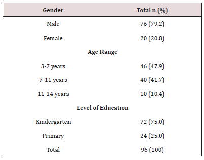
The sample consisted of ninety six children aged between 3 to 14 years. More than two thirds were male. Almost half 46 (47.9%) were in the 3–7year age group while a more than a third 40 (41.7%) were in the 11–14 year old age group. Children in Kindergarten were most represented (Table 1).
Table 1: Socio-Demographic Characteristics.

Distribution of Bacterial Plaque and Gingivitis

Large amounts of dental plaque deposits were found in half (45.21%) of children 3 to 7 years and 44.41% between the ages of 7 to11 years had.

Distribution of Dental Pathologies

Gingival bleeding was found in 59.19% of the 7–11 year old children, 55% in 3–7 year old’s and 16.14% in the 11–14 year old children. Two thirds (66.13%) of children aged between 7 and 11 years presented with dental caries Crown fracture (58.33%) was found in more than half of the children aged between 3 and 7 years. Overall the mean DMFT index was 2.76, the plaque index was 3.91 and the gingival bleeding index 2.32. Enamel opacities were found in 51.43% in children aged 3 to 7 years, 45.71% aged between 7 and 11 years and 2.86% in the 11–14 year old children). Enamel hypoplasia was identified in 42.86% in children aged 3 to 7 years, 50% in those between 7 and 11 years and 7.14% in the 11–14 year old children. Macrodontia was found in 71.43% in children aged between 11 and 14 years) compared to 28.57% in those aged between 7 and 11 years. Orthodontic problems like malocclusion and crowding was found in 9.37% of the children. The prevalence of trauma to dental soft tissues was 45.83% while the prevalence of dental abnormalities such was 31%. Nutritional practices, oral hygiene practices and oral health seeking behavior. More than two thirds (62.5%) of the children were exposed to cariogenic foods. Tooth brushing for the majority (70.83%) of the children was carried out by their mother. Half of the children brushed once a day. The majority brush their teeth before eating in the morning. Only 3 children brushed in morning and evening after meals. Two children reported brushing morning, afternoon and evening after meals. Only 13.54% had ever visited a dentist. Almost all 95.8% of the children had problems with word pronunciations. More than a third 38.5% of the children had difficulties talking aloud, a quarter had inconsistent speech (25%) and a quarter with incoherence when singings 20.8%. More than three quarters (80.2%) of the children presented with dental sensitivity (Table 2). Almost all 93.8% the children were not satisfied with their diet, 64.6% had difficulty with food intake (meals) and 62.5% avoid eating (Table 3).
Table 2: Functional Disorders.

Table 3: Dietary Behaviors.

Discussion

Autism spectrum disorders (ASDs) comprise a complex set of related developmental disorders that are characterized by impairments in communication, social interaction, and repetitive behaviors. Impairments in sensory processing are also very common. The prevalence of ASD is increasing and is currently estimated to affect 1 in 150 children worldwide [9]. It has been documented in many areas of the world that ASD is dominant among male children (3,4,5,6) as was found in the present study where there was a predominance of male children presenting with ASD. Various studies have reported the male: female ratio to range from 2:1 to 16:1 [9]. However, a recent systematic review carried out by Loomes R, in which fifty-four studies were analyzed, with 13,784,284 participants of whom 53,712 had ASD (43,972 boys and 9,740 girls) concluded that the true male-to-female ratio is not 4:1 as is often assumed but rather, it is closer to 3:1. There appears to be a diagnostic gender bias meaning that girls who meet criteria for ASD are at disproportionate risk of not receiving a clinical diagnosis [10]. Whatever the true ratio, clinical referrals to a specialist diagnostic centre have seen a steady increase in the number of girls and women referred. Because of the male gender bias, girls are less likely to be identified with ASD, even when their symptoms are equally severe. Many girls are never referred for diagnosis and are missed from the statistics. Emphasis is placed on the different manifestations of behavior in autism spectrum conditions as seen in girls and women compared with boys and men [7].
The age group most represented in the present study population was between 3 and 7 years old. This could be explained by the fact that after the appearance of the first symptoms during the first three years of life, either from birth or shortly after 12 months [3]. The kindergarten age group was more dominant in our study as three quarters of the children attended kindergarten. This age group is very important in children with ASD as an important transition period. The kindergarten transition is the first major educational transition in a child’s educational career. It is a particularly important transition for those with ASD because a successful kindergarten transition leads to better academic outcomes and better generalization of skills developed in the pre-school setting [8,9]. However, children with ASD begin with significant risk of an unsuccessful transition to kindergarten due to qualitative impairments in social communication and behavior. The use of recommended practices in kindergarten transition increases the chance of a successful transition for students with ASD [8,9]. Transitions in general, whether it be from one activity to another or one setting to another, are difficult for children with ASD [10]. These difficulties are often characterized by problems such as aggression, non-compliance, tantrums and a lack of consistent participation in activities [10]. It is because of these difficulties that the management of ASD is carried out using a multidisciplinary approach. The most effective strategies being based on educational programs [6,10]. In order to educate children with ASD, it is necessary to facilitate structured situations helping the patients to anticipate what is going to happen [6], since they show great fear and anxiety in the face of unknown situations [6], such as visiting the dental clinic. The dental care of these patients poses great difficulties, and in most cases, treatment is provided under general anaesthesia [6,11–16].

Eating Habits and Oral Hygiene of Children ASD

ASD are a major health and educational problem affecting many areas of daily living including eating. Children with ASD are often described as picky or selective eaters [12]. This was confirmed in the present study as two thirds of children preferred a soft and sweet diet. Poor reflexes during mastication and poor coordination of the tongue prevents normal swallowing and this results in a tendency to store food in the mouth and hence a preference for soft food. Various factors may contribute to food selectivity and several explanations have been proposed [12]. One of these factors relates to sensory sensitivity (also referred to as sensory defensiveness or sensory over-responsivity). Ayres [17] first described sensory defensiveness as the tactile domain (tactile defensiveness) in some children with learning and behavioral disorders. She described tactile defensiveness as an over-reaction to certain experiences of touch, often resulting in an observable aversion or negative behavioral response to certain tactile stimuli that most people would find innocuous. For example, children who show tactile defensiveness often have difficulty being cuddled and pull away from touch. It is possible that early tactile sensitivity may contribute to some of the sensory feeding issues such as difficulty with food textures seen in children with ASDs [12].

Oral Hygiene Status and Practice

The present study showed that the oral hygiene practices in terms of the frequency, duration and period of tooth brushing was very poor due to lack of co-operation, therefore the prevalence of dental caries and gingivitis is high. Parents and careers reported difficulties when they brushed the children’s teeth [14] further compounded by the lack of manual dexterity of autistic children. Morales-Chávez (2017) found a lack of cooperation in daily dental hygiene in 35.7% of children with ASD. Maintaining good dental hygiene is a difficult task for 40 to 50% of people with ASD [15]. Rada (2010) similarly argues that lack of oral hygiene is one of the biggest causes of rapid increases in caries rates and periodontal disease in young people with ASD compared to neurotypical children [16]. In general, children preferred soft and sweetened foods, and tend to ‘pouch’ food inside the mouth instead of swallowing it due to poor tongue coordination, thereby increasing the susceptibility to caries [18,19]. Studies have shown that children with autism exhibited a higher caries prevalence, poor oral hygiene and extensive unmet needs for dental treatment than non-autistic children [19–21] this concurs with the present study where high levels of dental plaque was found. The children also presented with a high mean DMFT index, a high plaque index and a high gingival bleeding index and therefore require stringent oral hygiene practices. In the present study, oral hygiene practices were carried out mainly by the mothers of the children.
Vajawat and Deepika [19] suggested that attempts should be made by parents, general dentists and periodontists to teach oral hygiene methods to the children by constant repetition and patience, as autistic individuals can develop skills over a period and lead a more productive and independent life [19]. Jaber MA [18] also suggested that oral health programmed that emphasize prevention should be considered of importance for children and young people with autism [18]. Another reason for high prevalence of oral diseases in autism children is poor access to oral health care facilities. In the present study 86.46% of the children with ASD never consulted a dentist and this could be due to the lack of infrastructure and human resources to manage the children in Cameroon. Bartolomé Villar and colleagues [22] in their review, reported that certain disorders are more prevalent in association with ASD — malocclusions, enamel hypoplasia, parafunctional habits (bruxism) and deleterious habits (mouth breathing, tongue thrusting, rumination) as well as a higher incidence of dental trauma, owing either to accidents or to self-imposed injuries [22]. Similarly, in the present study, dental malocclusion, enamel hypoplasia and enamel opacities were observed among the children with ASD, as was bad breath. Motta et al. [23] found a statistically significant association between halitosis and mouth breathing [23]). In the present study the prevalence of dental trauma to both the hard and soft tissues was very high as compared to the case-control study carried out by Altun et al. [24] on ASD children (AG) and a control group (CG). They reported that the rate of injury was higher among the AG (23%) than the CG (15%), though the difference between the 2 groups, however, was not statistically significant (P<.19). The most common type of dental injury was enamel fracture. The rate of enamel fracture was higher in the CG (59%) than in the AG (33%), and the distribution of types of traumatic injury differed significantly between the AG and CG (P>.01). According to their study, the maxillary central incisors were most affected and enamel fracture was mostly common. This is because the maxillary central incisors are the more prominent and are the first teeth receive impact. In our study, the high level of injuries might because of the play grounds, or the surroundings of the children are not protected from activities that might prevent impactful contacts. In conclusion, children with autism presented with a high prevalence of dental caries and periodontal diseases, bad breath, and dental trauma to hard and soft tissues.

Recommendation

Since autism patients present with many functional and clinical problems, a multidisciplinary approach is needed in Cameroon for the management of both dental and behavioral problems.

For more Lupine Publishers Open Access Journals Please visit our website: h
https://lupinepublishersgroup.com/
For more Journal of Otolaryngology-ENT Research articles Please Click Here:
https://lupinepublishers.com/otolaryngology-journal/
To Know More About Open Access Publishers Please Click on Lupine Publishers

Choanal Atresia Repair, A Comparison Between Transnasal Puncture With Dilatation And Stentless Endoscopic Transnasal Drilling

  Abstract Background: in this study we present the outcome of surgical repair of choanal atresia of 33 patients underwent t...