Showing posts with label Open Access Publishers. Show all posts

Friday, November 15, 2019

Lupine Publishers | Pre-cochlear Implantation Aural/Oral Rehabilitation, Is it Mandatory?

Lupine Publishers | Journal of Otolaryngology Research Impact Factor


Abstract

Background: The use of cochlear implantation (CI) can fully restore hearing. Consequently, speech production can improve over time and enters the normal rang when traditional amplification Devices (hearing aids) are unable to restore access to the full range of phonemic components of speech, a cochlear implant (CI) is a widely used treatment option for children with sensorineural hearing loss (SNHL).
Purpose: The aim of this study is to compare the functional benefit of the communicative skills of children with CI without pre-implantation aural/oral rehabilitation in relation to those with CI with pre-implantation 6 months aural/oral rehabilitation in order to compare the role of pre-implantation aural/oral rehabilitation on the communicative abilities of severe to profound and profound sensorineural hearing impaired children.
Method: This study has a prospective design. It started after final diagnosis and decision that all children are candidates for CI but half of them are fitted with behind the ear hearing aids and the other half of children are immediately implanted provided that the primary language assessment before rehabilitation is present in the medical files of these children. A 2nd language assessment was done after 12 months of language therapy to detect the progress of the language development. These sixty patients were divided into two groups:
a) Group A: Thirty children, who have used behind the ear hearing aids for one year before CI and attended regular language therapy.
b) Group B: Thirty children, who shifted immediately to cochlear implantation, and were enrolled in auditory training and language therapy for one year.
Results: Total language age of children using cochlear implant without pre-implantation aural/oral rehabilitation is significantly higher than that in the children while using hearing aids for one year before CI. Also, there is highly significant difference between frontal and back speech sounds in the children after immediate implantation with positive correlation.
Conclusion: Cochlear implant is safe & reliable technique. The fact that many profoundly hearing impaired children using immediate cochlear implant without pre-implantation aural/oral rehabilitation can develop functional levels of speech perception & production, develop competency level in a language other than their primary language and continuation of language therapy together with proper mapping accordingly is a must to enroll these children in main stream education.
Keywords: Hearing Aids; Cochlear Implant; Language; Speech Intelligibility Pre-implantation Rehabilitation
Abbreviations: SNHL: Sensorineural Hearing Loss; HA: Hearing Aids; CI: Cochlear Implantation

Introduction

Language in children begins to develop since birth and is nearly complete by the age of 6 years. Language skills, speech quality, expressive and receptive vocabulary are enhanced by exposure to aural language since as early an age as possible [1]. Children spend many hours in acoustic environments where target speech signals are embedded in competing sounds from multiple sources. In these environments, perception of target speech is assisted by a listener’s a listener’s ability to segregate the multitude of sounds into separate auditory streams, one cue to which is the angle of incidence of different sounds [2]. Children with profound sensorineural hearing loss (SNHL) experience delays in learning to understand the speech of others and to produce intelligible speech. There is solid evidence that moderate (or more severe) hearing impairment exerts a negative impact on speech, language, cognitive development, and early identification and management may be of great benefit to these children, through improved language, communication, mental health, and employment prospects [3]. The use of Hearing Aids (HA) or Cochlear Implantation (CI) can partially or fully restore hearing. Consequently, speech production can improve over time and enters the normal range. After hearing is restored, hearing impaired individuals use auditory feedback to adjust voice features such as voice intensity, intonation and vowel duration [4]. When traditional amplification devices (hearing aids) are unable to restore access to the full range of phonemic components of speech, a cochlear implant (CI) is a widely used treatment option for children with SNHL [5]. Cochlear Implants (CI) which are called as bionic ears are effective in trans- mitting salient features of speech, especially in quiet [6]. Because the goal of restored hearing in a deaf child is to enable useful hearing, a key measure of outcome should reflect how a deaf child’s experience with a CI develops into the effective use of spoken language. Parental surveys indicate that the outcome of their greatest concern after surgical intervention in children with SNHL is the level of spoken language achieved [7]. Cochlear implants have become a popular option for children with profound hearing loss. Evidence supporting the benefits of early implantation is found in experimental [1], developmental [2], and clinical cochlear implant studies [3]. The consensus is that children have the best opportunity to learn language during their first 5 years of life. According to [2], this critical period for language learning is particularly important in deaf and hearing-impaired children. Providing cochlear implants to deaf children at a young age may enable them to take advantage of this critical period for learning language and is likely to increase their chances for developing speech and language skills like those of normal-hearing children. Early implantation would also result in a decrease in the duration of auditory deprivation, a decrease considered to positively influence performance with a cochlear implant [4].

Objectives

The aim of this study is to compare the functional benefit of the communicative skills of children with immediate CI without preimplantation aural/oral rehabilitation in relation to those using hearing aid with pre-implantation aural/oral rehabilitation in order to compare the role of each amplification device and the effect of pre-implantation aural/oral rehabilitation on the communicative abilities of severe to profound and profound sensorineural hearing impaired children.

Subjects & Methods

This research was conducted during the period between the years 2017 and 2018. The study protocol was approved by the Otolaryngology Department Council of Beni-Suef University and Otolaryngology Department Council of King Abd Elaziz specialized hospital Jouf, Saudi Arabia. Consent to participate in this research was obtained from the subjects’ parents before commencement of the study. This study employed a comprehensive design to examine outcomes in multiple domains of communication in children who used either bilateral behind the ear hearing aids and preimplantation aural/oral rehabilitation or an immediate unilateral cochlear implant without pre-implantation rehabilitation for a period of one year. These were selected from children seeking language rehabilitation in Phoniatrics Unit, Beni-Suef University Hospital and children seeking language rehabilitation in Phoniatrics Clinic, King Abd Elaziz specialized hospital Jouf, Saudi Arabia. Shortly after confirmation of bilateral permanent hearing loss, thirty children were typically fitted with bilateral behind the ear hearing aids using the desired sensation level (DSL) prescription method and regularly attend aural/oral rehabilitation sessions. Thirty children underwent a comprehensive team evaluation for cochlear implant candidacy and received immediate unilateral cochlear implants without pre-implant aural/oral rehabilitation. All children received audiologic management and preschool rehabilitation and all children were enrolled in rehabilitation programs with a focus on the development of receptive &expressive language. Children were regular in Phoniatrics clinic, were asked to follow up auditory rehabilitation & language therapy program twice per week. Children with cochlear implants were followed every month for mapping of their speech processor and speech recognition testing. The study received ethical approval from the Hospital of Beni-Suef University and from King Abd Elaziz specialized hospital and written informed consent was obtained from all their parents. Collaboration between ENT clinic, Audiology clinic& Phoniatrics clinic was done in the form of ENT examination, audiological assessment, and language assessment and rehabilitation for all children. This study has a prospective design. It started after fitting half of the children with bilateral behind the ear hearing aids with aural/oral rehabilitation sessions and the other half of the children are immediately implanted, mapped, and regularly attended aural rehabilitation sessions provided that the primary language assessment before rehabilitate- ton is present in the medical files of all children. A 2nd language assessment was done after 12 months of language therapy to detect the progress of the language and the efficacy of pre-implantation aural/oral rehabilitation. These sixty patients were divided into two groups:
a) Group A: Thirty children, who have used behind the ear hearing aids for one year and attended regular language therapy despite those children, are candidates for cochlear implants.
b) Group B: Thirty children, who shifted immediately to cochlear implantation and had regular language therapy and were enrolled in auditory training.
Half patients were fitted with bilateral powerful digital signal processing BEHAs and used them for at least a 12-months period before CI. Hearing aid use was determined by parental and therapist reports. After surgical implantation of the device and an adequate healing period for the other half of the patients, the implants were activated (usually 4 weeks after surgery). The children were fitted with one of the two brands of speech processors using a behind the ear controller. Speech processors used in this study were OPUS 2 with standard Sonata electrode & Cochlear Freedom Processor with nucleus 24 k straight electrode.
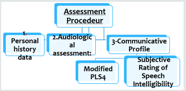
In this study using Modified Preschool Language Scale & Subjective Speech Intelligibility Test gave us a summary of the improvement of these children. This is matched with other studies which focused that both comprehension and expression of spoken language are important markers of parent-perceived success of a CI (Figure 1).
Figure 1
lupinepublishers-openaccess-journal-otolaryngology
Language Improvement Quotient: The language improvement quotient [8] was used to compare between the rates of progress in language in order to overcome the bias of age matching between the individuals in the study.
Language Improvement = 2nd language age -1st language age divided by duration of language rehabilitation.
a) A1 refers to language development of group (A) after using bilateral behind the ear hearing aids for 12 months which is calculated by this equation:
A1= 2nd language age -1st language age divided by 12(duration of rehabilitation)
b) B1 refers to language development of group (B) after using unilateral CI for 12 months which is calculated by this equation: B1= 2nd language age -1st language age divided by 12(duration of rehabilitation).
Speech analysis was performed using the Ain Shams assessment protocol which includes analysis of supra-segmental phonology (rate, stress and tonality), segmental phonology (consonants and vowels), nasal resonance and general intelligibility of speech. Assessment of auditory perception skills was performed evaluating a hierarchy of listening skills ranging from detection, to discrimination, identification, recognition and comprehension. Assessment of speech reading abilities was done and expressed as percent change over time.

Statistical Studies

Data was analyzed using SPSS, Statistical Package for the Social Sciences version 17 (SPSS Inc., Chicago, IL). Numerical data were expressed as mean, standard deviation, and range. For quantitative data, comparison was done using Mann-Whitney test (non-parametric t-test). A p-value < 0.05 was considered significant. Spearman-rho method was used to test correlation between numerical variables (r > 0.3 = no correlation, r = 0.3-0.5 = fair correlation, r = 0.5-0.1 = good correlation).

Results

Group (A) are hearing aids users for one year, Group (B) are CI users for 1 year. Demographic data of the 2 groups:
a) Age
b) Gender.
c) Incidence of hearing loss.
d) Psychometric evaluation.
e) Pure tone Audiometry.
f) First language age.
g) Radiology.
a) Age Distribution: Both groups are matched according to age. In group (A) the age of the children ranged between 3 years and 7years. In group (B). The age of the children ranged Between 3 years & 7 years, provided that all children were implanted before the age of 6 years.
b) Gender: No significant difference was noted in gender of both groups.
c) Incidence of Hearing Loss: In group (A) there were 24 children (80%) with congenital hearing impairment and 6 children (20%) with acquired hearing loss, while in group (B) there were 18 children (60%) with congenital hearing impairment and 12 children (40%) With acquired hearing loss.
d) Psychometric Evaluation: All children in group (A) had normal psychometric evaluation with a mean Value 87.5± 4.6; also, in group (B) all children had normal psychometric Evaluation with a mean value 86.4±5.1.
e) Pure Tone Results: Pure tone results of group (A) maintained a mean value of 27.9 dB HL. Group (B). Decreased in mean values from 65.7±8.2 dB HL. There was a highly significant difference (P=0.001) between group (A) and group (B) in favor of group (B).
f) First Language Age: Before start of therapy, both groups had no passive vocabulary and were Nonverbal. They used either babbling or vocal play.
g) Radiology: All Children both in groups (A) and (B) were having normal CT and MRI of Petrous bone.
Tables 1-3 demonstrate the progress of the language abilities, the auditory abilities and the speeding reading abilities of both groups, respectively, from the time just prior to the rehabilitation (either oral\ aural in group A or aural in group B) as compared to the evaluation done one year the rehabilitation (Table 4).
Table 1: Results of collective language improvement in both groups using paired-T test.
lupinepublishers-openaccess-journal-otolaryngology
Table 2: Results of progress in speech reading ability before and after rehabilitation in the 2 groups (using Paired-T test).
lupinepublishers-openaccess-journal-otolaryngology
Table 3: Difference in the speech ratings between the 2 groups after 3 years of rehabilitation (using Mann-Whitney test).
lupinepublishers-openaccess-journal-otolaryngology
Table 4: Results of collective language improvement in both groups using paired-T test.
lupinepublishers-openaccess-journal-otolaryngology

Discussion

The primary purpose of this study was to obtain comprehensive data on the development of language and speech skills in a group of permanent hearing impaired children. This group shared the common degree of bilateral hearing impairment (severe to profound or profound hearing impairment), they all sought amplification, and they all sought language rehabilitation after receiving amplification using primarily auditory-based cues. The study aimed also to investigate the difference between the language and speech development under two amplification conditions; bilateral behind the ear hearing aids and unilateral cochlear implants. The choice of language age deficit to compare language skills development among the studied groups is justified by the fact that three variables usually co-vary when language results are analyzed in children; age of use of the amplification device whether hearing aid (HAs) or cochlear implant (CI), the language age before start of rehabilitation, and the language age of children after the time of rehabilitation. The difference in ages at evaluation places the younger children at a maturational and developmental disadvantage in comparison with their older peers. Thus, analyzing the results in terms of language age scores might put the younger group at a disadvantage. At the same time, analyzing the results in terms of language age deficits, although more reasonable, but still, in theory, puts the older group at a disadvantage because of the impact of their ages giving higher values for the deficit from the scored language age. That’s why the hypotheses of using the language improvement quotient [8] after determining the exact language age, may be more realistic and less biased by the chronological age differences at the time of evaluation. In this study using Modified Preschool Language Scale & Subjective Speech Intelligibility Test gave us a summary of the improvement of these children. A perfect model for comparing the results of both devices may be practically impossible, given the current indications of cochlear implant use. In this study, which was applied on two groups with comparable ages, a comparison was made between the outcomes of the 2 devices along a period of (re)habilitation of one year in their course of therapy. If the CI group were doing better than the HA group, it would indicate that the selection criteria were too conservative and some of the HA users might be better off with a CI. This raises the suspicion of the fact that HA users plateau after a period of little progress or at least their progress continue at a less pace. Cochlear implants may have a superior effect on the acoustic environment of children more than hearing aids. During the 90s of the last centuries, and using the early models of speech processes, studies proved that CI users gained better results than HA users in language and perception skills [9-14]. The minimum age for implantation has progressively reduced [15]. Advantages of cochlear implants over hearing aids extended also the adult population [15]. In a study by [16], they found CIs and children with HAs, aged 4 to 5 years, differ significantly on language abilities and there were differences in articulation skills in favor of the CI users. Advances in sound processors and related software have enhanced the fidelity with which complex sounds are processed into physiologically meaningful codes [17]. This study pointed to the importance of conducting comprehensive assessments when evaluating whether a child with severe to profound sensory neural hearing loss would likely derive greater benefit from a cochlear implant compared to a hearing aid. To date, only a few systematic studies have involved large numbers of children who received implants at various ages and have investigated both the effects of age at implantation and the amount of experience with an implant. Most of such studies were concerned with the speech perception skills after cochlear implantation with a clear evidence of the effect of early implantation on rate of acquisition of such perception skills when they are implanted at 2 – 4 years of age [18]. Concerning the auditory abilities, the progress imposed by the effect of cochlear implantation group produced better abilities than the hearing aids group in the auditory abilities. This may be explained by the fact that the hearing aids group were more rigid to their habits of relying on their visual cues making the children less efficient in acquiring the training proficiency provided to them during therapy sessions. In an explanation of this, [19] described recruitment of the auditory cortex by the visual and somatosensory systems in congenitally deaf humans. They reported that the extent of crossmodal recruitment of the auditory cortex increases as the duration of deafness increases, deterring the restoration of auditory processing in the auditory cortex of long-term deafened individuals after cochlear implantation. They also suggested that the age beyond which the effects of cross-modal plasticity in the auditory cortex are more difficult to reverse is about 6.5 years. It has also been documented that there is a change in the cochlear place code during development [20]. This may be necessary for the formation of normal and effective connections between auditory centers and for the proper development of elements within the central auditory pathways. Early cochlear implantation may contribute to the maintenance of these important developmental milestones.

Conclusion

CI children showed better rate of language acquisition skills along a one-year use of the implant compared to a similar period of HA group of HA users. The implanted group demonstrated significantly better auditory abilities, better speech production skills, and better speech intelligibility one year after implantation and with aural rehabilitation - than the aided group with oral\aural rehabilitation. The implanted group also ended with significantly less or no speech reading abilities than the aided group one year after implantation. These results indicate the favorable effect implantation over the previous parameters. Language skills shows a significant difference between the two groups. Consequently, oral\aural rehabilitation with hearing aids – even for few monthsis not mandatory.
 
For more Lupine Publishers Open Access Journals Please visit our website: h
http://lupinepublishers.us/
For more Journal of Otolaryngology-ENT Research articles Please Click Here:
https://lupinepublishers.com/otolaryngology-journal/
 
To Know More About Open Access Publishers Please Click on Lupine Publishers


Saturday, October 26, 2019

Lupine Publishers | Occurance of Dizziness in patients with Tinnitus Complaint

Lupine Publishers | Journal of Otolaryngology

Abstract


Introduction: Dizziness is a regular complaint, often accompanied by other symptoms, especially tinnitus. Tinnitus is one of the three major otoneurological manifestations, alongside neurosensorial hearing loss and dizziness, being it, most of the times, the main complaint among patients. The relationship between the vestibular and cochlear system it’s rather known, many pathologies can originate from one of both systems.
Objective: Investigate the correlation between the symptoms of tinnitus and dizziness, analyzing the level of disturbance, the sensation of frequency (pitch) and intensity (loudness) of the tinnitus with dizziness complaint.
Methodology: A descriptive, observational and quantifying field study took place. 126 individuals with tinnitus complaint, from both sexes, were studied. The anamnesis was performed approaching audiological symptoms, the THI questionnaire was applied, as well as acuphenometry.
Results: 71 individuals (56,3%) referred to dizziness complaints associated with the tinnitus; women represented a larger number (41,3%) (p=0,017). In regards of the level of disturbance of the tinnitus, most of the patients 18,3% presented a low level, as for patients without dizziness 14,3% the quick level was present; the average Pitch is around 4.000 Hz in both groups, Loudness, on the other hand, was of 22 dBNS for individuals with dizziness and 26 dBNS for individuals without dizziness complaints. Conclusion: Meaningful results, regarding the relationship between tinnitus and dizziness, were not observed, therefore, it’s necessary to investigate if the tinnitus is from vestibular origin in order to seek improvements to the dizziness and thereafter, the tinnitus.
Keywords: Dizziness; Tinnitus; Audiology; Speech Therapy

Introduction

The corporal balance relies on the integrity of the vestibular system (labyrinth, nerve vestibulocochlear, cores, paths and interrelations of the central nervous system), somatosensorial system (receptors, sensors located on tendons, muscles and articulations) and vision [1]. The labyrinth is responsible for the balance and position of the body in location. Dizziness and/or imbalance cometo be when there is interference, both central and peripheral, in the regular operation of the body balance system. [2]. Dizziness can be defined, according to the Hearing and Balance Committee of the American Academy of Comitê de Audição e Equilíbrio da Academia Americana de Otolaryngology and Head/Neck Surgery [3], as every and any illusory feeling of motion without any real motion in relation to gravity. In the practice clinic, it’s one of the most frequent complaints. It affects between 20% and 30% of the general population, considering the epidemiological study of Neuhauser and collaborators [4]. In Brazil, an epidemiological study, performed in the city of São Paulo, showed that 42% of the individuals presented dizziness [5]. Occurrences can be found among any age range, from the first months of birth to the elderly population [6]. Causes can be many, such as: benign paroxysmal postural vertigo (VPPB), vestibular neuritis, Ménière’s disease, peristaltic fistula, circulatory, metabolic, hormonal or immunological, cervical spine changes, head trauma and psychoactive disorders, are one of the most common. This way, the symptoms coming from the dizziness may or may not originate from the vestibular system and comprises sensations described in many ways: vertigo (rotatory dizziness), imbalance, fluctuation or instability, presyncope our lipothymia, kinesis (motion sickness), oscillopsia, falling [7]. Dizziness of nonvestibular origin are often ill-defined, most of the times they are labeled as uneasiness, light-headedness, sensation of fainting. Also, in rare cases, they might represent symptoms of the central nervous system and/or may be associated with exclusively ocular disorders, ischemic episodes, metabolic disorder, neurological, cardiac or cervical diseases [8]. However, dizziness is usually due to primary or secondary functional disorders of the vestibular system, it might be classified as rotatory (vertigo – when the illusion of motion has rotatory characteristics) or non-rotatory (when the illusion of motion has no rotatory characteristics). Regular vertigo is more common among peripheral syndromes rather than central ones. The peripheral vertigo is usually aggravated by eye shutting, unlike what happens with central vertigo. Both peripheral and central vertigo can be unleashed or worsen with head motion (this being the most common kind of rotatory dizziness). On the other hand, non-rotatory dizziness may be oscillating, hesitant, fluctuance, wavering, among others. In order to determine the vestibular source an examination of the alterations of the vestibular system is required [9]. Dizziness is usually followed by other symptoms; it’s intensity can cause loss of balance and falls. Normally, the dizziness appearance is accompanied by neurovegetative symptoms, megrims, eyesight darkening and lack of concentration.
Dizziness is also highly associated with auditory symptoms, such as hearing loss, sensation of auricular plenitude and, mainly, tinnitus [10,11]. The tinnitus is one of the 3 major otoneurological manifestations, alongside neurosensorial hearing loss and dizziness, being it, most of the times, the main complaint among patients, especially elder ones [12,13]. The tinnitus, also known as tinnitus, can be defined as auditory illusion, in other words, an endogenous sound illusion, not related to any outside source of stimulation [14]. The presence of tinnitus might be a factor of great negative repercussion in one’s life, jeopardizing sleep, concentration during day-to-day and professional, as well as social life. Many times, it affects the emotional balance of the patient, unleashing or worsening states of depression and anxiety [15]. A study performed in the city of são Paulo shows that 22% (430 individuals) present tinnitus [16]. The tinnitus is as symptom that can be caused by a number of medical conditions: otological affections, neurological, cardiovascular, metabolic, pharmacologic, odontological, psychologic, side effects of medications and possible drug ingestion, such as caffeine, nicotine and alcohol [17]. Up to date theories to explain the source of tinnitus defend the hypothesis that it occurs due to anomalous and spontaneous neural activity in the central pathways, auditory or not, being a consequence of sensory deprivation, aftermath of cochlear lesion [18,19]. The description of the tinnitus’s characteristics might vary from patient to patient, from “pure tone” sound to a “whistle”, a “noise” or even a “whisper”, etc. Perceivable in one or both ears, and yet in the head, with no specific side. It can be constant or intermittent, being absent for some periods of time. Its intensity may vary from light to very intense [18]. Patients with dizziness, resistant to various treatments, can be as hard to conduct as patients with high level of tilllindus disturbance, these that might happen simultaneously or independently. Both dizziness and noise are extremely common symptoms at the practice clinic, as shown by the study performed by Moreira and collaborators [20] where out of 27 individuals affected by dizziness, 16 (59,2%) also complained about noise.
The relationship between the vestibular and cochlear systems is rather known. Many pathologies may originate in one of those systems or simultaneously, as well as having one of them as primary source due to influence in other systems functionality. Therefore, it’s possible that changes in the posterior labyrinth (Semicircular Canal) fluids may cause tinnitus [21]. It’s noticeable that both disorders jeopardize the individual’s quality of life. The tinnitus may affect one’s sleep, concentration, emotional balance and social life. On the other hand, dizziness, apart from other mentioned symptoms, might hinder the individual’s performance during activities that require quick head motions and, also, tasks that imply flexing the torso and the head [22]. Innumerable reports of dizziness among patients with tinnitus complaints were observed in a Multidisciplinary service of attendance to patients with tinnitus. The aforementioned research main goal was to investigate the correlation between dizziness and tinnitus symptoms on those patients, analyzing the level of disturbance, the sensation of frequency (pitch) and intensity (loudness) of tinnitus with dizziness complaint.

Material and Methods

The present study was performed in one School Clinic of the Speech Therapy Course located in João Pessoa. 126 individuals with tinnitus complaint were evaluated, 81 females and 45 males, ages ranging from 17 to 83 years old, all attended in the Multidisciplinary Tinnitus Service. It was a descriptive research. As for technical procedures, a field study is more fitting, as it tries to deepen between the dizziness and its relationship towards tinnitus. In order to validate these hypotheses, a transversal and observational study, of quantitative characteristics, was performed. In accordance with the 466/12 Resolution of the National HealthCommittee, referring to ethics regarding research that involves human subjects, the study was approved by the Ethics in Research with Human Subjects Committee (prot. N. 0129/12). Clearance was obtained through the signing of the Term of Free and Enlightened- TCL by volunteers and/or responsible for the project. All patients were subjected to the following procedures:
I. Answering anamnesis questions which emphasize the patient’s auditory symptoms, in order to collect personal data of the individual; data about the tilllindus - location of the tinnitus (right, left, in the head or undetermined), time of onset, how it came to be (gradual, sudden, after noise exposure, other), kind (continuous, pulsatile, intermittent), characteristics (whistle, rain, noise, waterfall, bee, other); and other possible associated symptoms, such as dizziness.
II. In order to obtain more directed information regarding the patient’s tinnitus, the Tinnitus Handicap Inventory (THI) questionnaire was applied, as an interview. The THI is a questionnaire that evaluates the severity of the tinnitus, with 25 questions approaching three dimensions: nine questions related to emotional aspects (frustration, anger, irritability, anxiety, depression and insecurity); eleven questions related to functional aspects (stress, concentration, sleep, workplace interference, house responsibilities and social activities); antivideos socials); and five questions related to the catastrophic aspects (despair, lack of self-control, inability of acceptance, perception of terrible illness) (Lim et al, 2010). Those 25 questions allow the following possibilities of answer: “yes”, “no” and “sometimes”, each having a score of “4 points”, “0 points” and “2 points”, respectively. This said, each question will add points to its category, be it functional, emotional or catastrophic, and the total sum, varying from 0 to a 100. Depending on the result, the level of disturbance caused by the tinnitus might be classified as, by the total sum, as:
a) LEVEL 1 (Quick): Score 0 - 16. Only perceived in quiet environments.
b) LEVEL 2 (Light): Score 18 - 36. Easily masked by environmental and easily forgotten during day-to-day activities.
c) LEVEL 3 (Moderated): Score 38 - 56. Perceived in the presence of background noise, however, day-to-day activities can still be performed.
d) LEVEL 4 (Severe): Score 58 - 76. Almost always perceived, leads to disturbance in sleep patterns and may interfere in daily activities.
e) LEVEL 5 (Catastrófico): Score 78 - 100. Always perceived, sleep patterns disturbances, difficulty performing daily activities.
Later on, acuphenometry was performed in order to obtain data regarding the sensation of intensity (loudness) and frequency (pitch) of the tinnitus. The test took place in an acoustic cabin, using the audiometer model AVS 500, of the Vibrasom brand. To unilateral tinnitus, the sound was supplied to the contralateral ear and, if bilateral, to the ear with best hearing [23]. In order to identify what kind of tinnitus, the Pure Continuous Tone, Pulsatile Pure Tone and Modulated Frequency were presented, in audible intensity, so that the patient could choose which resembles his own tinnitus more. To measure Pitch, the chosen tone was presented in frequencies ranging from 125 to 8000 Hz. And to investigate loudness, the same tone was presented in the frequency identified by the patient, with audible intensity, incrementing 1 db.

Data Analysis

The data was registered in an Excel spreadsheet for further analysis. Initially, a static descriptive analysis was performed, in order to verify the frequency of the variables studied (tinnitus, dizziness, age, gender).
As it follows, the inferential static analysis was also performed, with the help of adequate tests, in order to verify:
a) The correlation between variables: Spearman Correlation test aiming to verify the level of relationship between pairs of variables of interest, such as THI x Dizziness, Gender x Dizziness.
b) Comparison between pitch and loudness values to each group, with or without dizziness: parametric test t of Student to independent samples of interval variables of normal distribution; or its nonparametric correspondent when needed.
c) The differences were considered meaningful when p0,05 was presented. The static analysis was performed through the Software Statistical Package for Social Sciences (SPSS), version 20.0.

Results

Out of the 126 evaluated individuals (64.3%) are female and 45 (35.7%) male, with average age of 49,33 years. All of them presented tinnitus, whom 71 (56,3%) also complain about dizziness, whereas 55 (43,7%) don’t. Based on this, it was noticed that most part of the studied population stated complaints regarding tinnitus associated dizziness, also the static analysis shows that the variable gender has great interference in the presence or absence of dizziness. Regarding the THI questionnaire, the patients with dizziness presented bigger results for the light level, however patients without dizziness, presented bigger results to the quick level. As for tinnitus characteristics, both groups presented average pitch around 4.000 Hz, with no meaningful statistical difference between them. On the other hand, loudness average of the tinnitus was 22 dBNS for the dizziness group and 26 dBNS for the individuals without dizziness, no meaningful difference between the groups was found (Tables 1-3).
Table 1: Descriptive and inferential statistics of the variables gender and dizziness.
lupinepublishers-openaccess-journal-otolaryngology
Meaningful difference (p<0,05) according to Spearman’s correlation test.
Table 2: Descriptive and inferential statistics of tinnitus disturbance and dizziness.
lupinepublishers-openaccess-journal-otolaryngology
Meaningful difference (p<0,05) according to Spearman’s correlation test.
Table 3: Averages of pitch and loudness of the tinnitus.
lupinepublishers-openaccess-journal-otolaryngology
Meaningful differences when p<0,05 according to the t Student test.

Discussion

Both tinnitus and dizziness are otoneurological complaints often presented at the practice clinic. Patients that exhibit tinnitus might also show signs of alteration and/or vestibular complaints. In this research, was observed that most of the population filed dizziness complaints. This data corroborates with literature, seeing it as complaint usually reported alongside tinnitus [20,21]. Data from table one shows that more women (41.3%) presented complaints about dizziness associated with tinnitus and there was a meaningful correlation (p=0,017), in accordance with what literature shows [24-27]. It is believed that the higher occurrence among females might be due to factors such as: variation of the hormonal cycle, higher occurrence of migraines and the fact that women are more likely to seek medical attention [27,28]. Therefore, the variable gender directly affects the presence or not of dizziness. The disturbance caused by the tinnitus may vary greatly, and there are factors that appear to be associated with a higher level of disturbance, such as stress, psychiatric disorders and gender [29,30]. The THI analysis, expressed in table 2, showed that most of the patients with dizziness presented light level 18,3%, whereas, patients without dizziness presented the quick level 14,3%. Therefore, the data is compatible with the ones presented by the studies of Xavier [31] and Lim et al. [32] that highlight the light and quick level as the most common among the samples of tinnitus. It can also be observed in the present study that, despite the variables THI and Dizziness lack of meaningfulness (p= 0,71), patients with dizziness complaints seem to exhibit a higher level of disturbance regarding tinnitus than the ones without complaint. In regards to the characteristics of the tinnitus evaluated by acuphenometry (Table 3), when compared to frequency values, both groups showed an average pitch around 4.000 Hz, in other words, the two groups, regardless dizziness complaints, exhibited the pitch of the tinnitus in acute frequencies, with no meaningful statistical difference between them (p=0,105). These findings are in accordance with the studies of Urnad and Tochetto [33] and Suzuki and collaborators [34] which also verifies values referring to the tinnitus’s pitch in acute frequencies.
This is strongly related with the fact that most patients with tinnitus present hearing loss in these frequencies. The researchers state that there is a connection between the tinnitus’s pitch and the region of the frequency of the maximum hearing loss. Taking intensity into consideration, the average loudness of the tinnitus was 22 dBNS for the group with dizziness and 26 dBNS for individuals without it, no verified meaningfulness between the two groups (p=0,234). This data goes against the values of the studies of Buzo and Carvallo [35] and Tugumia and collaborators [36] that verified loudness varying from 5 to 15 dBNS, this way, our population shows a bigger sensation of intensity to the tinnitus. Meaningful results regarding the relation between tinnitus and dizziness were not observed in this sample. Probably, the dizziness complaints of these patients are not related to the vestibular system and, consequently, to the tinnitus. Understanding that the auditory and vestibular systems are intimately related, it becomes necessary that the patient with dizziness complaints be directed to and otoneurological evaluation, in order to investigate if the source of dizziness is vestibular, what could possibly strengthen the relationship between the tinnitus and the dizziness. Afterwards, it is possible to seek treatment in order to improve both conditions, simultaneously, as it is for vestibular rehabilitation which is used as a therapeutic process for dizziness when associated with tinnitus, possibly decreasing the level of disturbance caused by the tinnitus, as in accordance with the study presented by Zeigelboim and collaborators [37].

Conclusion

Based on the results found in the researched sample:
a) Most of the patients with tinnitus presented dizziness [38].
b) The variable female gender showed meaningfulness regarding the presence of dizziness.
c) An average pitch of around 4.000 Hz was found for individuals with and without dizziness.
d) Average loudness was 22 dBNS for individuals with dizziness and 26 dBNS for individuals without.
e) The level of disturbance of the tinnitus showed no meaningfulness/relationship with dizziness.

For more Lupine Publishers Open Access Journals Please visit our website: h
http://lupinepublishers.us/
For more Journal of Otolaryngology-ENT Research articles Please Click Here:
https://lupinepublishers.com/otolaryngology-journal/

To Know More About Open Access Publishers Please Click on Lupine Publishers 

Saturday, September 28, 2019

Lupine Publishers | Radiofrequency Ablation for Snoring and Sleep Apnoea

Lupine Publishers | Journal of Otolaryngology
 

Abstract

Radio frequency proves to be a useful tool for snoring/ sleep Apnoea cases. Its advantage includes relative precision in incision making, relative bloodless fields if used appropriately, decrease postoperative pain and excellent healing with fibrosis which aids in stiffening tissues. Radiofrequency is high frequency alternating current used to ablate (cut/coagulate) tissues. It can be applied to nasal turbinate’s, soft palate, tongue base, tonsils etc. and it can be used to perform various procedures in the cutting mode to improve obstructive sleep disordered breathing. The objective/aim was to assess efficacy of radiofrequency as a tool for procedures/surgeries for snoring/ sleep apnoea. The parameters assessed were post-op pain, post- op blood loss, reduction in subjective snoring sounds by patients and partner, reduction in AHI post operatively.

Methods

The procedures were carried out over a period of three years. All cases that came to us had complaints of snoring, difficulty in breathing and sleep disturbances at the hospital departments were included in the study [1]. A total of 25 cases were studied. A thorough history, clinical examination in all and flexible endoscopy /sleep study were carried out according to the case. The radiofrequency SUTTER BM 7180 machine was used to treat patients. The power settings used were from 2 - 6 in the cutting and coagulation mode. The procedures were carried out under; local or general anaesthesia with oral intubation and a throat pack.

RF Tonsillectomy

Exposing the tonsil on either side, the To-bite radiofrequency forceps or the RF needle was used to incise /open the plane for tonsillar dissection. Dissection was carried out with the same achieving haemostasis at the same time. If properly done bleeding was minimal and pain scores were low post operatively. Fossa deepened and stiffened post operatively. RF setting of 2-3 in cutting mode and 5-6 in coagulation mode was used.

RF Adenoidectomy

Can be performed after retracting lower edge of the palate with tongue depressors or tourniquets and coagulating the adenoid with bipolar forceps, the lower edge of the adenoid can be dissected using RF needle or ball point. Bleeding is negligible and wound heals well. There was no case of postoperative haemorrhage. Ideal for recurrent adenoids. RF setting of 5-6 in the coagulation mode.

RF Palate

It is temperature controlled RF volumetric reduction of the palate in order to stiffen or scar the soft palate. The Sutter RF bipolar probe is used to deliver energy to the soft palate at various points. Blanching has to be avoided. The subsequent stiffening occurs over 6 weeks. It was done under local anaesthesia as an outpatient procedure with no bleeding and low pain scores. Subjective decrease in snoring was achieved even in one sitting.

RF Tongue Base

It is temperature controlled volumetric tongue base reduction by giving RF energy to multiple sites of post tongue base with Sutter RF bipolar forceps [2]. Three sittings of reduction gave a significant reduction in tongue base tissue. There was no incidence of tongue base oedema or infection. The procedure could be done under local or general anaesthesia.

RF UP3

It is achieved by uvular and periuvular lateral cuts and trimming of lower edge soft palate with rf in cutting mode and subsequent suturing and tonsillectomy with pillar suturing. The postoperative widening contracture /stiffening helps in achieving a good result.

RAUP

For snoring is done by uvular and lateral cuts and redefining the post pillars. Tonsillectomy may be combined. It achieves its result due to removal of the redundant mucosa and subsequent healing with fibrosis. Subjective decrease in snoring is achieved by most patients. RF is used in the cutting mode.

Result (Table 1)

Table 1:
lupinepublishers-openaccess-journal-otolaryngology

Discussion

Of the 27 patients who underwent treatment with radiofrequency, of the 5 palate cases 2 patients got a pain score of 4 and 3 patients 0-1. RAUP patients had a varied score of 1 to 4. RF adenoidectomy was relatively pain free and tonsillectomy was between 4-5. Rf tongue base had very low pain scores. There was no postoperative bleeding in any of the cases. Intra operative bleeding was encountered in tonsillectomy when rf was used in the cutting mode. RF Palate in one sitting can give a reduction in snoring by 50-70%. Rf in cutting mode if used inappropriately can give rise to bleeding issue otherwise not.

Conclusion

Rf appears to be an efficient tool for snoring/sleep apnoea procedures because of:
a) Ability to cut fast and maintain a relatively bloodless field.
b) Ability to cut and coagulate at various settings.
c) Decrease intraoperative blood loss.
d) Induces fibrosis and stiffening of tissues.
e) Decrease postoperative pain.

Other Advantages

a) The instrument /unit appears dynamic with a good unique feel.
b) Procedures can be performed under local / gen. Anaesthesia.
c) Instruments are autoclavable/recurring cost is lower.
d) Machine is ambulatory.
e) Minimally invasive.

For more Lupine Publishers Open Access Journals Please visit our website: h
http://lupinepublishers.us/
For more Journal of Otolaryngology-ENT Research articles Please Click Here:
https://lupinepublishers.com/otolaryngology-journal/
To Know More About Open Access Publishers Please Click on Lupine Publishers

Follow on Linkedin : https://www.linkedin.com/company/lupinepublishers
Follow on Twitter   :  https://twitter.com/lupine_online 

Saturday, August 24, 2019

Lupine Publishers | Updates about Vertebrobasilar Insufficiency in Dizziness

Lupine Publishers | Journal of Otolaryngology


Abstract

Vertebrobasilar insufficiency (VBI) is a result of transitory ischemia of the vertebrobasilar arterial system (VBS) that can produce a variety of symptoms that are on their own are ambiguous. Symptoms include dizziness, vertigo, lightheadedness, headaches, visual changes, diplopia, ataxia, weakness in limbs, pain and stiffness of the neck. Vestibular and visual symptoms can arise suddenly and dissipate rapidly as well, all while preceding more serious symptoms like stroke and death. There are a variety of tests that audiologists and physical therapists can perform as screeners for this impairment, but imaging is an essential component of the diagnosis. Neuroimaging with angiography, magnetic resonance angiography (MRA), magnetic resonance imaging (MRI), and transracial Doppler tests are commonly used. Diagnosis and treating VBI requires a multidisciplinary and interdisciplinary approach. VBI has been documented to be exaggerated and over diagnosed in part due to the vague and transient nature of the symptomology. This paper will further outline the anatomy of the VBS, symptoms of VBI, risk factors, and diagnostic criteria.
Keywords: Atherosclerosis; Balance; Dizziness; Vertebrobasilar Arterial System; Vertebrobasilar Insufficiency

Introduction

The vertebrobasilar arterial system (VBS) supplies the vestibular and cochlear nuclei and is comprised of the vertebral artery (VA), basilar artery (BA), anterior-inferior cerebellar artery (AICA), and the posterior-inferior cerebellar artery (PICA) [1]. The VA and BA supply blood to the pons, medulla, cerebellum, mesencephalon, thalamus, occipital lobes, and of course, the peripheral and central vestibular system, as mentioned above [2]. The reason why changes in the VBS have a greater effect on the vestibular system rather than affecting both cochlear and vestibular function is that the cochlear system also received blood flow from the carotid artery which protects it from suffering the same impairments as the vestibular system which only received blood from the labyrinthine branches of the vertebrobasilar arteries [3]. Furthermore, the ischemia of the VBS can affect both peripheral and central vestibular structures by causing isolated dizziness attacks resulting from ischemia to the vestibular nuclei and/or vestibular cochlear nerve, or directly affecting the labyrinth [3]. The typical cause of hemodynamic changes in this system are the result of atherosclerosis; but other causes are embolism, and arterial dissection, or rarely migraine, fibromuscular dysplasia, and coagulopathies [3]. Considering the anatomy and physiology, it is easy to understand how changes or limitation in blood flow can induce disequilibrium, vertigo, and/or vision changes. Symptoms include dizziness, vertigo, nystagmus, imbalance, lightheadedness, headaches, mental confusion, aural fullness, tinnitus, hearing changes, nausea, vomiting, syncope, diplopia, blurred vision, blindness, ataxia, difficulty swallowing, dysarthria, pain and stiffness in neck or shoulder, and weakness of the extremities [1–4]. While it is possible for isolated and sudden attacks of vertigo to occur, vertigo alone is does not meet the diagnostic criteria for VBI [2,3]. Visual disturbances are commonly congruent with vertigo in VBI patients, but in fact, visual symptoms such as diplopia, visual hallucinations, changes in visual field, and blindness are more common than vertigo [3]. Patient report visual changes, diplopia, palsy of the oculomotor nerve, and seeing spots [2,3]. The onset of symptoms occurs rapidly and can be described as an attack by occurring during the change of position or suddenly reaching maximal affect within 5 minutes of the start and lasting anywhere from 2 minutes to 30 minutes commonly (but has been reported up to 24 hours) [4]. The resolution of the symptoms (or attack) occurs quickly. The frequency and antecedent of symptoms vary making it additionally difficult to diagnose based on patient complaints alone. The incidence is difficult to calculate as it has been recorded that VBI diagnosis has been exaggerated and inappropriately used to diagnose other conditions [5]. There is however a sex effect in that VBI is more likely to be found in men (rather than women) after the fourth decade of life [3]. 

Discussion

Differential diagnosis of VBI required careful examination to separate the symptoms from other conditions. Recording a detailed case history, performing the appropriate clinical measures, and having the essential imaging is required for adequate differential diagnosis. Case history will be necessary to identify possible risk factors such as: hyperlipidemia, hypertension, cerebrovascular diseases, carotid disease, heart disease, smoking, alcoholism, diabetes, and hyperglycemia [2,5]. Record and description of total symptoms at time of attack and clarifying any vagueness of response or other possible related incidences will be helpful. Also recognize that position changes like moving from laying/sitting to standing can induce symptoms. A vestibular assessment may be useful in differentiating the symptoms from other disorders. In the vestibular assessment the clinician may find that a patient with VBI and vestibular decruitment and hyperactive caloric responses [2,4]. Although hyperactive caloric responses may be observed, the two results of this test, canal paresis and directional preponderance, do not distinguish between an intracranial lesion or a labyrinthine impairment; however, the presence of decruitment/and or hyperactivity is indicative of an impairment as normal patients do not present with these clinical findings [4]. Some patients were also found to have abnormal function in the optokinetic pattern test, eye tracking, or visual suppression test in the videonystagmography (VNG) [1]. Another assessment conducted by the audiologist that may be useful in the Auditory Brainstem Response (ABR) when the ischemic is in the AICA displaying a substantial increase in interpeak latency between waves I-IV and II-IV [1].
A retrospective study done in China by Hu et al. [5] found that out of 773 patients diagnosed with VBI only 67 (8.67%) of them had true VBI. Other conditions to consider are benign paroxysmal positional vertigo (BPPV), Meniere’s disease, vestibular neuronitis, syncope, heart disease, abnormal blood pressure, sudden deafness, infectious diseases, and brain trauma [5]. Accordingly, neuroimaging will be useful in differential diagnosis. Tests include angiography, magnetic resonance angiography (MRA), magnetic resonance imaging (MRI), transracial Doppler, angio-tomography, and cardiologic studies [3,4]. Arteriography is highly useful in diagnosing VBI, but fewer patients are willing to have this test completed due to the risks of arterial catheters, low blood flow, or stroke [4]. The non-invasive test of MRA can be completed to identify possible occlusion or stenosis in neck or intracranial vessels [1,3]. This test has already been useful by identifying that the proximal regions of the vertebral arteries is the location of highest incidence causing VBI, as recorded with MRA [3]. When investigating the basilar artery, the MRA and angio-tomography are found to have similar sensitivity and specificity. The use of transcranial Doppler test is a low-cost, noninvasive, and pain-free test that is used to measure the speed and direction of intracranial arterial blood flow [3]. The plasticity index (PI) recording from the Doppler test is useful in predicting early hemodynamic intracranial variations; however, there is a sex effect in that speech and PI results are opposing for men and women as they age [3]. As evident with the above explanation, there are many useful tests that can be completed to aid in diagnosis of VBI and the need to multidisciplinary and interdisciplinary team is essential.

Conclusion

Understanding risk factors and possible symptoms is helpful in differential diagnosis, however, the risks and symptoms are broad and vague. Patients may present with the exact same symptomology but have varying diagnosis. The use of objective measures, such as caloric testing, and neuroimaging will be vital to accurately diagnosis. Due to the vast symptoms and fluctuating presentations, there is no set diagnostic criteria that can be applied to all patients. This is a concern to protect patients from the progression of the impairment which are stroke and possibly death. Correspondingly, more research needs to be conducted to understand the progression of stenosis as it gradually worsens over time.

For more Lupine Publishers Open Access Journals Please visit our website:
https://lupinepublishers.us/
For more Journal of Otolaryngology-ENT Research articles Please Click Here:
https://lupinepublishers.com/otolaryngology-journal/

To Know More About Open Access Publishers Please Click on Lupine Publishers

Choanal Atresia Repair, A Comparison Between Transnasal Puncture With Dilatation And Stentless Endoscopic Transnasal Drilling

  Abstract Background: in this study we present the outcome of surgical repair of choanal atresia of 33 patients underwent t...