Showing posts with label lupine publishers indexing journals. Show all posts

Thursday, August 15, 2019

Lupine Publishers | Apnea, Hypopnea and Their Individual Effects on Daytime Sleepiness and Sleep Quality

Lupine Publishers | Journal of Otolaryngology

Abstract

Background: Obstructive sleep apnea and hypopnea syndrome (OSA) is defined as a reduction or cessation of the airflow in the human airway. It effects nearly 18 million Americans and weight gain is the main predisposing factor. In this study, we aimed to investigate the effects of apnea and hypopnea individually.
Material and Methods: 83 participants were included in the study and they are divided into two groups as apnea predominant or hypopnea predominant. Pittsburg quality of sleep index (PQSI) and Epworth sleepiness scale (ESS) are completed for all subjects and full-night attended polysomnographic evaluations are done.
Results: ANOVA test was used to compare the inter-group variances. Between the two study groups, no statistical significance was reported between the PSQI or ESS scores.
Conclusion: The effects of apnea and hypopnea are similar on sleep quality or day-time sleepiness, however further studies also investigating the duration of the events as well are needed.
Abbreviations: OSA: Obstructive Sleep Apnea/Hypopnea Syndrome; PSG: Polysomnography; PSQI: Pittsburgh Sleep Quality Index; ESS: Epworth Sleepiness Scale.

Introduction

Obstructive sleep apnea and hypopnea syndrome (OSA) is reviewed under the sleep related breathing disorders. The diagnostic criteria must satisfy daytime sleepiness, fatigue or nonrestorative sleep, waking up with breath holding, gasping or choking, witnessed apnea periods and comorbidities such as hypertension, mood disorder, cognitive dysfunction, coronary artery disease or type 2 diabetes mellitus may accompany the disease. The full-night polisomnography (PSG) must demonstrate at least five obstructive respiratory events (apnea, hypopnea, respiratory effort related arousals) per hour of sleep (apnea/hypopnea index, AHI), but AHI below 15 needs the abovementioned signs for a complete diagnosis [1]. OSA may be seen in any age group, nevertheless published data from several countries indicate that OSA associated with daytime sleepiness occurs in 3% to 7% of adult men and 2% to 5% of adult women. However, because many individuals with OSA do not endorse daytime sleepiness, the prevalence of the disease is likely much higher [2]. The major predisposing factor for OSA is excess body weight. It has been estimated that nearly 60% of moderate to severe OSA is attributable to obesity. The risk of OSA increases as the degree of additional weight increases, with an extremely high prevalence of OSA in people with morbid obesity [3]. Several factors are implicated in the development of OSA [4].
The main cause addresses the reduction of the expansion forces of the dilator muscles of the upper airways. The capacity of the muscles decreases more during the REM sleep. Additional factors are excessive or elongated tissues of the soft palate, macroglossia, tonsillar hypertrophy, and a redundant pharyngeal mucosa [5]. OSA and comorbidities such as stroke, hypertension, metabolic syndrome, cardiovascular diseases or endocrinologic disorders are well taught in years, however the individual effects of apnea or hypopnea alone are never considered. To best of our knowledge, the published data does not mention which entity alone is more harmful to systemic functions or at least sleep, apnea or hypopnea? The Pittsburgh Sleep Quality Index (PSQI) is an effective instrument used to measure the quality of sleep in the adult. It differentiates “poor” from “good” sleep by measuring seven domains: subjective sleep quality, sleep latency, sleep duration, habitual sleep efficiency, sleep disturbances, use of sleep medication, and daytime dysfunction over the last month. A global sum of “5” or greater indicates a “poor” sleeper [6]. The Epworth Sleepiness Scale (ESS) is a self-administered questionnaire with 8 questions. Respondents are asked to rate, on a 4-point scale, their usual chances of dozing off or falling asleep while engaged in eight different activities. The higher the ESS score, the higher that person’s daytime sleepiness and scores higher than 10 are significant. In this study, we compared apnea versus hypopnea due to the sleep quality and daytime sleepiness individually.

Materials and Methods

Between July 2017 and January 2019, 327 cases complaining of snoring, daytime sleepiness and witnessed sleep apnea periods were referred to full-night polisomnography. 150 cases were diagnosed as OSA were enrolled in the study. After the final assessment, a total number of 83 participants were chosen. The ethical committee approval was taken from Okmeydani Training and Research Hospital (48670771–514.10) and informed consent are taken from all participants.

PSG

3 Channel EEG (F4-M1, C4-M1, O2-M1), 2 channel EOG, chin, right and left tibialis anterior EMG, body position sensor, oro-nasal thermal sensor, nasal pressure sensor, thoracic and abdominal sensors, ECG, pulse-oximetry and synchronous video recordings and breath sound recordings were the parameters recorded through the night. The examination, sleep and wake periods and sleep related disorders were scored according to the criteria of the American Academy of Sleep Medicine [7].

Clinical Examination and Laboratory Tests

All participants underwent a detailed otolaryngologic examination including the fiberoptic naso-pharyngolaryngoscopy. Mueller maneuver was made to detect the pharyngeal collapse, vallecula epiglottica was visualized to assess the bulkiness, Friedmann Tongue Positions and Tonsil Gradings are made to determine the glossopharyngeal patency. Complete blood count and routine biochemical blood tests including the thyroid function tests were studied. PSQI and ESS were completed for each participant.

Study Design

In order to emphasize the individual effects of apnea or hypopnea, each PSG were examined and if apnea was higher than hypopnea by 50% or vice versa, that participant was included in the study. The study group, therefore, was divided into two as apnea predominant (AP) and hypopnea predominant (HP). Comorbid pulmonary or neurologic disorders, sleep disorders other that OSA (Central sleep apnea, Hypersomnolence, Parasomnias, Circadian rhythm disorders, etc.) were excluded. Also, pediatric population were not included in the study.

Statistical Analysis

A statistical analysis was performed using IBM SPSS Statistics 22 (IBM SPSS, Turkey). Continuous data was displayed as the mean ± standard deviation. Statistical significance was a p-value of greater than 0.05. A Shapiro–Wilk test showed the normal distribution of the parameters. ANOVA test was used to compare the normally distributed inter-group comparisons of the descriptive statistical methods (mean, standard deviation, and frequency) and the quantitative data.

Results

A total number of 83 participants aged between 24–64 years (mean 46±10 years) were studied. 61 were male (73.49%) and 22 were female (26.51%). The inter-group characteristics are shown in Table 1. The PSQI between AP and HP groups were not statistically significant (p=0.205). Similarly, ESS between AP and HP groups were also not statistically significant (p=0.240) (Figure 1).
Figure 1: AP versus HP by means of PSQI and ESS.
Table 1: Subject characteristics and statistical comparisons.
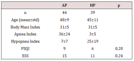
ANOVA test showed no statistical significance among variances.
OSA is defined as the reduction or cessation of airflow for at least ten seconds. The entity is almost every time in association with snoring, and between the snores, airflow may stop completely (apnea) or reduction in the airflow (hypopnea) may happen. If there is a body effort to breathe, the disease is termed obstructive, otherwise it is central. In the presence of a collapsible airway, sleepinduced loss of tonic input to the upper airway dilator muscle motor neurons allows the pharyngeal airway to collapse [8]. The general reaction to this airway obstruction is arousal; sleep then resumes, leading to repeated cycling of sleep, intermittent hypoxia, and arousal throughout the night. Neurocognitive effects of OSA include daytime sleepiness and impaired memory and concentration; cognitive impairment and neural injury may develop in association with sleep apnea [9,10]. Sleep-disordered breathing and OSA are not reported frequently in animals but a natural animal model

Discussion

OSA is defined as the reduction or cessation of airflow for at least ten seconds. The entity is almost every time in association with snoring, and between the snores, airflow may stop completely (apnea) or reduction in the airflow (hypopnea) may happen. If there is a body effort to breathe, the disease is termed obstructive, otherwise it is central. In the presence of a collapsible airway, sleepinduced loss of tonic input to the upper airway dilator muscle motor neurons allows the pharyngeal airway to collapse [8]. The general reaction to this airway obstruction is arousal; sleep then resumes, leading to repeated cycling of sleep, intermittent hypoxia, and arousal throughout the night. Neurocognitive effects of OSA include daytime sleepiness and impaired memory and concentration; cognitive impairment and neural injury may develop in association with sleep apnea [9,10]. Sleep-disordered breathing and OSA are not reported frequently in animals but a natural animal model of OSA is English bulldogs, which have been used to study upper airway anatomy and physiology and the pharmacologic treatment of OSA. English bulldogs have an enlarged soft palate and narrow oropharynx and display many of the clinical features of OSA, including snoring, sleep-disordered breathing, oxyhemoglobin desaturation during sleep, frequent arousal from sleep, and hypersomnolence with shortened sleep latencies [11]. OSA in English bulldogs is not related to obesity, as it often is in humans. OSA has been modeled in a variety of species by using surgical tracheostomy and subsequent intermittent occlusion of the endotracheal tube [12]. Schoorlemmer et al. [13] produced obstructive apnea in conscious rats by using an inflatable balloon implanted in the trachea and apneic episodes of as long as 16 s in duration could be created during sleep. However, animal models of intermittent hypoxemia have several drawbacks. In many cases, the models mimic severe human OSA and may be less applicable to most clinical OSA. In addition, animals exposed to intermittent hypoxemia develop hypocapnia, whereas human OSA is characterized by hypercapnia. Furthermore, human OSA typically is associated with obesity, which is not always considered in animal studies. In addition, OSA causes sleep fragmentation, which may have independent effects on metabolism. Thus, exposure of animals to intermittent hypoxemia produces repeated arousals and changes in sleep architecture that are comparable to those in clinical OSA, yet the effects may not be persistent, limiting their use for studying long-term metabolic consequences of OSA [14].
Animal models are troublesome to study the long-term effects of sleep fragmentation. The sleep quality is a bio-psycho-social parameter that may never be evaluated in animal models; for example, as we refer to excessive daytime sleepiness, the subject is asked whether the sleepiness occurs in the passive state such as resting periods or during the active periods such as work-time or social interactions. Moreover, the sleep architecture, ultradian rhythm or sleep quality may not be assessed in animal models. In our study, the effects of apnea or hypopnea on sleep quality or daytime sleepiness did not differ. This might have several reasons; first of all, all PSG were done elsewhere, our otolaryngology clinic is not capable to perform full-night attended PSG. This situation has some major drawbacks; it is impossible to mark each breath disorders epoch by epoch on the screen of the test computer, but we rather have a brief report of the night. This makes it impossible to calculate the duration of the respiratory events. Therefore, we only could compare the nature of the events by their scores or numbers (Table 1). Secondly, there are some other factors that may interfere with the sleep architecture such as drops in the oxygen levels; not every subject has the same decreasement in their oxyhemoglobine when they have the same level and duration of airway collapse. Thirdly, limb movements also disrupt the sleep quality and impede normal daytime cognitive functions. Finally, arousals are another issue to study; if OSA is recently developed in the subject, the peripheric chemoreceptors detecting the airflow cessation are more sensitive and arousal happens imminently, if the disease is longer the receptors may become insensitive that happens in sleep continuity despite the airflow cessation. Nonetheless, to best of our knowledge, there is no other study that tried to investigate the effects of apnea of hypopnea individually on sleep quality or daytime sleepiness.

Conclusion

The effects of apnea and hypopnea are similar on sleep quality or daytime sleepiness, however further studies also investigating the duration of the events as well are needed.

For more Lupine Publishers Open Access Journals Please visit our website: h
https://lupinepublishersgroup.com/
For more Journal of Otolaryngology-ENT Research articles Please Click Here:
https://lupinepublishers.com/otolaryngology-journal/

To Know More About Open Access Publishers Please Click on Lupine Publishers

Tuesday, August 13, 2019

Lupine Publishers | Aging, Hearing Loss and Tinnitus

Lupine Publishers | Journal of Otolaryngology

Abstract

This article explores the occurrence of tinnitus and hearing loss in the elderly. Both are highly prevalent after 60 years old. It highlights the relationship between hearing loss, tinnitus and aging. And it points out some possible form of intervention.

Introduction

Since the end of the century, the number of elderly people worldwide has increased. By the year 2015 there will be about 2 billion people over the age of 60. This population growth is probably due to the decline in the birth rate, improvement of socio-economic conditions of life and progress of modern medicine [1]. However, increasing longevity does not characterize good population health. As life expectancy progresses, there is also a greater number of chronic and incapacitating diseases, which must be properly monitored since they can cause problems for the quality of life of this population. Decreased functional capacity of organs and tissues is the main feature of aging and carries an increased risk of chronic degenerative diseases and declining status of the elderly in the family and in society. The gradual loss of physical energy and economic productivity tends to isolate and deprive of sources of information and communication [2].
However, some physiological changes that happen with the advancing age will only manifest itself from the third age. For example, the human ear reaches maturity around 18-20 years and from this age the hearing organ begins to age, either by loss of sensory cells, neurological degeneration, exposure to ototoxic agents or noise [3]. Hearing loss in the elderly, also known as presbycusis, is a bilateral hearing loss for high frequency sounds. The main clinical manifestations of presbycusis include symmetrical and slowly developing sensorineural hearing loss, high pitched tinnitus and speech recognition disorders [4]. In the case of presbycusis, generally, the hearing thresholds increase significantly between 70 and 80 years of age and reach another stable stage at high levels after 80 years of age, especially in high frequencies [5]. Tinnitus is a common complaint defined as a sound in the head or ears that occurs in the absence of any external acoustical source [6]. It may be caused by several conditions: otological, metabolic, neurological, orthopedic, cardiovascular, pharmacological, odontological and psychological, which in turn may be present concomitantly in the same individual [7]. It affects about 15% of the world population. It can occur at any stage of life, but the highest prevalence occurs in the elderly, probably due to deterioration of the auditory and vestibular systems [8,9]. Tinnitus is the second most common otorhinolaryngological complaint in the elderly [10], with tinnitus often more disturbing than hearing loss [11]. Approximately 33% of the elderly population is affected by tinnitus and 15% to 25% of them present interference with the quality of life caused by this symptom [12]. Both hearing loss and tinnitus can trigger important communication problems, which in turn lead to difficulties in social, occupational and family adaptation. It is very common for elderly individuals to report that they can hear but not understand speech. Some studies have attempted to identify the relationship between age, gender, hearing loss and tinnitus [13-15]; however, which not confirm that tinnitus discomfort could be explained by age, gender, and hearing loss. The hearing loss might be the most dangerous factor and if the its serious, the incidence of the tinnitus became higher. So, tinnitus in the elderly may be the result of a combination of factors. Therefore, other issues are likely to be investigated, such as psychological issues or underlying diseases. To date, is known that exist a high prevalence of hearing loss and tinnitus in the elderly, and that these have a high impact on the patient’s quality of life. Becoming a factor of great negative repercussion for this population, hindering sleep, social life, concentration on daily and professional activities. The first step in care is to investigate the patient’s history. A detailed anamnesis, which should address, in addition to questions about tinnitus, associated diseases, patient’s lifestyle, diet, genetics, general health and the current effects of the disease on the patient’s life. In addition to the anamnesis, the use of questionnaires is important in the evaluation of individuals with tinnitus, as it helps to confirm the presence of tinnitus and determines the severity of the symptoms [16,17]. Treatment may be based on direct reduction of severity or elimination of tinnitus, such as working with the patient’s emotions in the face of tinnitus [18]. Other therapies also known are relaxation techniques, cognitive-behavioral therapy (CBT), psychological counseling, sound therapy, including hearing aids or sound generators, or a combination of these approaches [19]. Until now, CBT is the oneoff have scientific evidence for tinnitus treatment. However, when the patient has hearing loss and tinnitus, the Tinnitus Retraining Therapy has been demonstrating that this model of intervention becomes a treatment option for the relief of tinnitus in the elderly people [20] and of hearing aids could reduce the perception of tinnitus sound intensity and the bothersome with this symptom and with hearing loss [21].

Conclusion

In this way, neither hearing loss or tinnitus should be neglected, it is important found the better management and intervention that relieve the symptom of tinnitus and hearing loss. This can improve elderly quality of life.

For more Lupine Publishers Open Access Journals Please visit our website: h
https://lupinepublishersgroup.com/
For more Journal of Otolaryngology-ENT Research articles Please Click Here:
https://lupinepublishers.com/otolaryngology-journal/

To Know More About Open Access Publishers Please Click on Lupine Publishers

Wednesday, July 24, 2019

Lupine Publishers | Oral Health Status in Children with Autism Spectrum Disorder Presenting in Yaoundé, Cameroon

Lupine Publishers | Journal of Otolaryngology

Abstract

Introduction: Children with Autism Spectrum Disorders (ASD) present with a lifelong neuro-developmental disorder that makes them prone to oral diseases. The aim of the present study was to describe the oral health status of children with ASD attending autism centers of Yaoundé, Cameroon.
Methodology: A descriptive cross-sectional study was conducted in seven specialized centers caring for children with autism. A structured questionnaire and clinical examinations were used to collect the data.
Results: Ninety six children aged between 3 to 14 years, the majority of whom were boys (80%), were recruited into the study. The majority were from kindergarten (75%) and the remainder primary schools. Tooth brushing for most of the children (71%) was carried out by their mother. Half of the children brushed once a day with the help of their mothers usually in the morning before breakfast, using toothbrushes and toothpaste. Most of the children consumed soft and sweet diets that included sweetened fruit juices and had never been to a dentist. The prevalence of caries was 66.13%. Dental caries was more pronounced in the 7 to 11 year old age group. Almost half (45.21%) had gingivitis associated with plaque and calculus and 59.19% had gingival bleeding. The prevalence of trauma to both hard and soft oral tissues was 45.83% while the prevalence of dental abnormalities was 31%. The treatment needs of the children include dental fillings, scaling and curettage.
Conclusion: Children with ASD presented with a high prevalence of dental caries and periodontal diseases, bad breath and dental trauma to hard and soft tissues.
Recommendation: Since autism patients present with many functional and clinical problems, a multidisciplinary approach is needed in Cameroon for the management of medical, dental and behavioral problems.
Keywords Autism Spectrum Disease; Cameroon; Children; Oral health

Introduction

Oral diseases such a dental caries and periodontal disease are the most common chronic diseases and are important public health problems because of their high prevalence [1]. Oral health and general health are interlinked, affecting and affected by one another. General health can affect oral health. Medical conditions often have oral implications and consequences [1,2] and one disease affecting general health that can affect oral health is Autism which is considered a disease of special care within the oral health care services. Results of the 1994–1995 National Health Interview Survey of the USA on access to care and use of services indicated that the most prevalent unmet health need among children with special health care needs was oral health care [3]. During the 2014– 2016 survey, the prevalence of children aged 3–17 years who had ever been diagnosed with a developmental disability increased from 5.76% to 6.99%. During this same time, the prevalence of diagnosed autism spectrum disorder and intellectual disability did not change significantly. The prevalence of autism spectrum disorder, intellectual disability, other developmental delay, and any developmental disability was higher among boys when compared with girls [4]. Autism spectrum disorder (ASD) is a persistent neurodevelopmental condition with early childhood onset. It begins in early childhood and persists throughout adulthood affecting three crucial areas of development: communication, social interaction, and creative or imaginative play [5]. The universal occurrence of autism spectrum disorders (ASD) was investigated about twenty-six years ago and it was thought to occur only in western industrialized countries that had high levels of technological development.
Over the years, knowledge about ASD and its prevalence has been documented as being on the rise in different regions of the world, with most literature coming from the western world, while the situation in Africa remains unclear [5]. No specific oral manifestations of ASD have been described, although the oral hygiene is known to be deficient [6]. Nevertheless, many authors have found the prevalence of caries and of periodontal disease to be no different compared with non-autistic individuals [6] and in some cases the prevalence of caries in children with ASD may even be comparatively lower [6]. Between 60–95% of all patients with ASD have an unusual sensory profile, including dysfunction in registering oral sensitivity. Studies have shown a high prevalence of periodontal and orthodontic problems such as crowding of the dentition in children with autism as compared to non-autistic children [7,8]. In addition, systemic medications taken by autistic patients may also affect their oral health. It has been reported that nearly 25% of young people with ASD present with gingival hyperplasia related to antiepileptic medications. Furthermore, some medication may compromise the health and function of the oral environment causing decreased salivary flow, dry mouth and a reduced mechanical and antibacterial action of saliva [8]. In recent years, there has been an increasing awareness of autism and autism-related diseases in the Cameroon. However, there is a paucity of information regarding the oral manifestations and the oral health care needs of children with ASD. The aim of the present study was to determine the prevalence of the oral manifestations in autism patients and to establish their oral health needs.

Methodology

This descriptive study was carried out in Yaoundé between January to July 2017 in all the specialized centers taking care of children with autism. These centers currently take care of children with ASD aged between 3–14 years regardless of gender. The eight centers identified were in five districts of the city of Yaoundé. Outside the Center for Special Education for children with ASD located in the district of Yaoundé I, the target population consisted of 145 children in four districts of the city of Yaoundé (II, IV, V, and VI). The districts of Yaoundé III and VII lacked education centers. Of the eight identified centers, seven agreed to participate in the study from the 4 districts based on the approval or consent of parents or guardians. Children aged between three and fourteen years old were included in the study. The inclusion criteria for participation in the study were defined before the sample were screened and consisted of the following: a diagnosis of ASD, the understanding of very simple instructions and written informed consent from the parents or caregivers. Data collection began following the approval by the relevant authorities as well as the consent from the parents and caregivers of the participants. Information from the patients was collected using a data capture sheet after which clinical examinations were carried out on a mobile dental clinic under bright natural light. The parents / caregivers of the participants received an explanation of the study, together with an information sheet and informed consent form. Those who agreed to participate completed a questionnaire evaluating the socio-demographic status of the children, medical history, functional disabilities, nutritional status, nutritional habits and oral hygiene of the patients. Data not adequately recorded were obtained by interviewing the parents or caregivers.
A single dentist was used to carry all the clinical dental examinations using a mirror and probe on a portable dental chair and guidable light source of enough power for adequate intraoral exploration. At the end of the examination, the parents and caregivers received a report on the oral disorders found, with suggestions for referral regarding the necessary treatments and dental care. Clinical examinations were carried out to evaluate the oral health status, the prevalence of dental caries, periodontal diseases, enamel hypoplasia, hard and soft tissues trauma and other pathologies in the oral cavity. The DMFT index was determined assessing the decayed, missing and filled teeth. Oral hygiene was rated using the Simplified Oral Hygiene Index (OHI-S). The criteria of this index are related to the sum of the Plaque Index Score (PI-S) and the Calculus Index Score (CI-S). In both cases the surfaces of index teeth were examined: vestibular surfaces of 11, 16, 26 and 31; lingual surfaces of 36 and 46. In the absence of index teeth, the entire sextant was recorded to the maximum degree (except for the third molars).

Statistical Analysis

Data were captured into Microsoft excel 2010 and exported to SPSS for statistical analysis. Results were presented in the form of tables using Microsoft Excel 2010. Bivariate analysis was carried out using Chi-square test for qualitative variables and ANOVA for quantitative variables p= 0.05.

Ethical Considerations

Approval to carry out this study was obtained from the Ethics Committee of the University des Montagnes. Informed consent was obtained from parents and all information obtained was strictly confidential. Participants were informed that they were free to leave the study at any stage without penalty to their further management.

Results

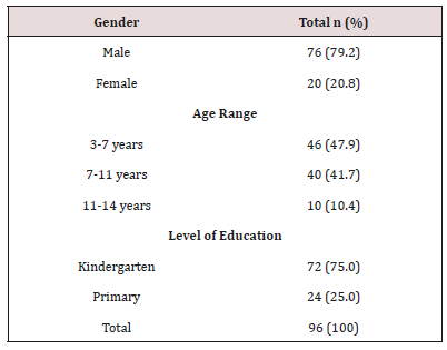
The sample consisted of ninety six children aged between 3 to 14 years. More than two thirds were male. Almost half 46 (47.9%) were in the 3–7year age group while a more than a third 40 (41.7%) were in the 11–14 year old age group. Children in Kindergarten were most represented (Table 1).
Table 1: Socio-Demographic Characteristics.

Distribution of Bacterial Plaque and Gingivitis

Large amounts of dental plaque deposits were found in half (45.21%) of children 3 to 7 years and 44.41% between the ages of 7 to11 years had.

Distribution of Dental Pathologies

Gingival bleeding was found in 59.19% of the 7–11 year old children, 55% in 3–7 year old’s and 16.14% in the 11–14 year old children. Two thirds (66.13%) of children aged between 7 and 11 years presented with dental caries Crown fracture (58.33%) was found in more than half of the children aged between 3 and 7 years. Overall the mean DMFT index was 2.76, the plaque index was 3.91 and the gingival bleeding index 2.32. Enamel opacities were found in 51.43% in children aged 3 to 7 years, 45.71% aged between 7 and 11 years and 2.86% in the 11–14 year old children). Enamel hypoplasia was identified in 42.86% in children aged 3 to 7 years, 50% in those between 7 and 11 years and 7.14% in the 11–14 year old children. Macrodontia was found in 71.43% in children aged between 11 and 14 years) compared to 28.57% in those aged between 7 and 11 years. Orthodontic problems like malocclusion and crowding was found in 9.37% of the children. The prevalence of trauma to dental soft tissues was 45.83% while the prevalence of dental abnormalities such was 31%. Nutritional practices, oral hygiene practices and oral health seeking behavior. More than two thirds (62.5%) of the children were exposed to cariogenic foods. Tooth brushing for the majority (70.83%) of the children was carried out by their mother. Half of the children brushed once a day. The majority brush their teeth before eating in the morning. Only 3 children brushed in morning and evening after meals. Two children reported brushing morning, afternoon and evening after meals. Only 13.54% had ever visited a dentist. Almost all 95.8% of the children had problems with word pronunciations. More than a third 38.5% of the children had difficulties talking aloud, a quarter had inconsistent speech (25%) and a quarter with incoherence when singings 20.8%. More than three quarters (80.2%) of the children presented with dental sensitivity (Table 2). Almost all 93.8% the children were not satisfied with their diet, 64.6% had difficulty with food intake (meals) and 62.5% avoid eating (Table 3).
Table 2: Functional Disorders.

Table 3: Dietary Behaviors.

Discussion

Autism spectrum disorders (ASDs) comprise a complex set of related developmental disorders that are characterized by impairments in communication, social interaction, and repetitive behaviors. Impairments in sensory processing are also very common. The prevalence of ASD is increasing and is currently estimated to affect 1 in 150 children worldwide [9]. It has been documented in many areas of the world that ASD is dominant among male children (3,4,5,6) as was found in the present study where there was a predominance of male children presenting with ASD. Various studies have reported the male: female ratio to range from 2:1 to 16:1 [9]. However, a recent systematic review carried out by Loomes R, in which fifty-four studies were analyzed, with 13,784,284 participants of whom 53,712 had ASD (43,972 boys and 9,740 girls) concluded that the true male-to-female ratio is not 4:1 as is often assumed but rather, it is closer to 3:1. There appears to be a diagnostic gender bias meaning that girls who meet criteria for ASD are at disproportionate risk of not receiving a clinical diagnosis [10]. Whatever the true ratio, clinical referrals to a specialist diagnostic centre have seen a steady increase in the number of girls and women referred. Because of the male gender bias, girls are less likely to be identified with ASD, even when their symptoms are equally severe. Many girls are never referred for diagnosis and are missed from the statistics. Emphasis is placed on the different manifestations of behavior in autism spectrum conditions as seen in girls and women compared with boys and men [7].
The age group most represented in the present study population was between 3 and 7 years old. This could be explained by the fact that after the appearance of the first symptoms during the first three years of life, either from birth or shortly after 12 months [3]. The kindergarten age group was more dominant in our study as three quarters of the children attended kindergarten. This age group is very important in children with ASD as an important transition period. The kindergarten transition is the first major educational transition in a child’s educational career. It is a particularly important transition for those with ASD because a successful kindergarten transition leads to better academic outcomes and better generalization of skills developed in the pre-school setting [8,9]. However, children with ASD begin with significant risk of an unsuccessful transition to kindergarten due to qualitative impairments in social communication and behavior. The use of recommended practices in kindergarten transition increases the chance of a successful transition for students with ASD [8,9]. Transitions in general, whether it be from one activity to another or one setting to another, are difficult for children with ASD [10]. These difficulties are often characterized by problems such as aggression, non-compliance, tantrums and a lack of consistent participation in activities [10]. It is because of these difficulties that the management of ASD is carried out using a multidisciplinary approach. The most effective strategies being based on educational programs [6,10]. In order to educate children with ASD, it is necessary to facilitate structured situations helping the patients to anticipate what is going to happen [6], since they show great fear and anxiety in the face of unknown situations [6], such as visiting the dental clinic. The dental care of these patients poses great difficulties, and in most cases, treatment is provided under general anaesthesia [6,11–16].

Eating Habits and Oral Hygiene of Children ASD

ASD are a major health and educational problem affecting many areas of daily living including eating. Children with ASD are often described as picky or selective eaters [12]. This was confirmed in the present study as two thirds of children preferred a soft and sweet diet. Poor reflexes during mastication and poor coordination of the tongue prevents normal swallowing and this results in a tendency to store food in the mouth and hence a preference for soft food. Various factors may contribute to food selectivity and several explanations have been proposed [12]. One of these factors relates to sensory sensitivity (also referred to as sensory defensiveness or sensory over-responsivity). Ayres [17] first described sensory defensiveness as the tactile domain (tactile defensiveness) in some children with learning and behavioral disorders. She described tactile defensiveness as an over-reaction to certain experiences of touch, often resulting in an observable aversion or negative behavioral response to certain tactile stimuli that most people would find innocuous. For example, children who show tactile defensiveness often have difficulty being cuddled and pull away from touch. It is possible that early tactile sensitivity may contribute to some of the sensory feeding issues such as difficulty with food textures seen in children with ASDs [12].

Oral Hygiene Status and Practice

The present study showed that the oral hygiene practices in terms of the frequency, duration and period of tooth brushing was very poor due to lack of co-operation, therefore the prevalence of dental caries and gingivitis is high. Parents and careers reported difficulties when they brushed the children’s teeth [14] further compounded by the lack of manual dexterity of autistic children. Morales-Chávez (2017) found a lack of cooperation in daily dental hygiene in 35.7% of children with ASD. Maintaining good dental hygiene is a difficult task for 40 to 50% of people with ASD [15]. Rada (2010) similarly argues that lack of oral hygiene is one of the biggest causes of rapid increases in caries rates and periodontal disease in young people with ASD compared to neurotypical children [16]. In general, children preferred soft and sweetened foods, and tend to ‘pouch’ food inside the mouth instead of swallowing it due to poor tongue coordination, thereby increasing the susceptibility to caries [18,19]. Studies have shown that children with autism exhibited a higher caries prevalence, poor oral hygiene and extensive unmet needs for dental treatment than non-autistic children [19–21] this concurs with the present study where high levels of dental plaque was found. The children also presented with a high mean DMFT index, a high plaque index and a high gingival bleeding index and therefore require stringent oral hygiene practices. In the present study, oral hygiene practices were carried out mainly by the mothers of the children.
Vajawat and Deepika [19] suggested that attempts should be made by parents, general dentists and periodontists to teach oral hygiene methods to the children by constant repetition and patience, as autistic individuals can develop skills over a period and lead a more productive and independent life [19]. Jaber MA [18] also suggested that oral health programmed that emphasize prevention should be considered of importance for children and young people with autism [18]. Another reason for high prevalence of oral diseases in autism children is poor access to oral health care facilities. In the present study 86.46% of the children with ASD never consulted a dentist and this could be due to the lack of infrastructure and human resources to manage the children in Cameroon. Bartolomé Villar and colleagues [22] in their review, reported that certain disorders are more prevalent in association with ASD — malocclusions, enamel hypoplasia, parafunctional habits (bruxism) and deleterious habits (mouth breathing, tongue thrusting, rumination) as well as a higher incidence of dental trauma, owing either to accidents or to self-imposed injuries [22]. Similarly, in the present study, dental malocclusion, enamel hypoplasia and enamel opacities were observed among the children with ASD, as was bad breath. Motta et al. [23] found a statistically significant association between halitosis and mouth breathing [23]). In the present study the prevalence of dental trauma to both the hard and soft tissues was very high as compared to the case-control study carried out by Altun et al. [24] on ASD children (AG) and a control group (CG). They reported that the rate of injury was higher among the AG (23%) than the CG (15%), though the difference between the 2 groups, however, was not statistically significant (P<.19). The most common type of dental injury was enamel fracture. The rate of enamel fracture was higher in the CG (59%) than in the AG (33%), and the distribution of types of traumatic injury differed significantly between the AG and CG (P>.01). According to their study, the maxillary central incisors were most affected and enamel fracture was mostly common. This is because the maxillary central incisors are the more prominent and are the first teeth receive impact. In our study, the high level of injuries might because of the play grounds, or the surroundings of the children are not protected from activities that might prevent impactful contacts. In conclusion, children with autism presented with a high prevalence of dental caries and periodontal diseases, bad breath, and dental trauma to hard and soft tissues.

Recommendation

Since autism patients present with many functional and clinical problems, a multidisciplinary approach is needed in Cameroon for the management of both dental and behavioral problems.

For more Lupine Publishers Open Access Journals Please visit our website: h
https://lupinepublishersgroup.com/
For more Journal of Otolaryngology-ENT Research articles Please Click Here:
https://lupinepublishers.com/otolaryngology-journal/

To Know More About Open Access Publishers Please Click on Lupine Publishers
Choanal Atresia Repair, A Comparison Between Transnasal Puncture With Dilatation And Stentless Endoscopic Transnasal Drilling

  Abstract Background: in this study we present the outcome of surgical repair of choanal atresia of 33 patients underwent t...