Showing posts with label Journal of Otolaryngology-ENT Research. Show all posts

Friday, September 30, 2022

Primary Laryngeal Lymphoma and Voice Hoarseness: A Rare Etiology for A Common Clinical Feature


Abstract

Primary Laryngeal Lymphoma is rare both as a hematological and as a laryngeal malignancy. Unlike squamous cell carcinomas, lymphomas require chemo-radiotherapy as 1st-line treatment. A 67-year-old lady presented with voice hoarseness, dyspnea and dysphagia for solids. An otherwise unremarkable physical showed “hot-potato like” voice. Labs were normal, while laryngoscopy and CT revealed an epiglottic mass, which was resected. Biopsy results showed DLBCL. The patient then underwent chemotherapy with complete success. Voice hoarseness has a vast differential diagnosis depending on its character and complementary symptoms. Less than a 100 cases of Primary Laryngeal Lymphoma have been reported in the literature, proving the rarity of this clinical entity.

Keywords:Primary laryngeal lymphoma; voice hoarseness; diffuse large B-cell lymphoma; larynx; lymphoma; epiglottis; dysphagia; dyspnea; “hot-potato like” voice

Abbreviations:CT: Computed Tomography; PET-CT: Positron Emission Tomography- Computed Tomography; NHL: Non-Hodgkin Lymphoma; ECOG: Eastern Cooperative Oncology Group; WBC: White Blood Cells; NEU: Neutrophils; LYM: Lymphocytes; MONO: Monocytes; EOS: Eosinophils; BASO: Basophils; RBC: Red Blood Cells; Hgb: Hemoglobin; Hct: Hematocrit; MCV: Mean Corpuscular Volume; MCH: Mean Corpuscular Hemoglobin; PLT: Platelets; ESR: Erythrocyte Sedimentation Rate; LFTs: Liver Function Tests; AST: Aspartate Transaminase; ALT: Alanine Transaminase; γ-GT: Gamma-glutamyl transferase; Bil: Bilirubin; dBil: Direct bilirubin; iBIl: Indirect bilirubin; Cr: Creatinine; BUN: Blood Urea Nitrogen; Glu: Glucose; Alb: Albumin; TC: Total Cholesterol; TGL: Triglycerides; LDH: Lactate Dehydrogenase; CRP: C reactive protein; DLBCL: Diffuse large B-cell lymphoma; LPR: Laryngopharyngeal Reflux; NK: Natural Killer Cells; R-CHOP: Rituximab, Cyclophosphamide, Andriamycin, Oncovin, Prednisone; HD-MTX: High Dose Methotrexate; CNS: Central nervous system; SCC: Squamous Cell Carcinoma; MALT: Mucosa-Associated Lymphoid Tissue; EBV: Eibstein Barr Virus; BMI: Body Mass Index

Read More About This Article Click on Below Link:
https://lupinepublishers.com/otolaryngology-journal/fulltext/primary-laryngeal-lymphoma-and-voice-hoarseness.ID.000222.php

Read More About Lupine Publishers Google Scholar Articles:
https://scholar.google.com/citations?view_op=view_citation&hl=en&user=dMOUw-wAAAAJ&cstart=20&pagesize=80&citation_for_view=dMOUw-wAAAAJ:EBV337fEn3EC


Friday, July 29, 2022

Trazodone Not Only A Stabilizer in Allergic Rhinitis


Letter to Editor

We report the case of a 44 years old woman referred to the outpatient Sleep Disorder Service of the Neurological Clinic, Marche Polytechnic University for a symptomatology characterized by difficulty in maintaining sleep and a feeling of unrefreshing sleep on waking. On clinical history, she referred an allergic rhinitis, treated with short cycles of inalatory steroids and vasoconstrictors. At the moment of our observation, she did not assume any treatment [1]. Due to the reported symptoms, particular attention was paid to the possible presence of sleep disturbances, in particular sleep-related breathing disorders. In this respect, patient reported a morning dry mouth. Further, the partner referred an important snoring and continuous lightening of sleep. Nocturnal apnea events were not reported as well as discomfort or abnormal movements in the legs. No other disturbance suggestive for NREM or REM sleep disturbances, nor fewer specific phases were evidenced. Patient was then submitted to a polygraphy which showed a rhonchopathy associated with air flow limitations, a high arousal index and elevated heart rate variability. The exam excluded periodic limb movements (Figure 1) [2,3]. Considering the history of allergic rhinitis and also to evaluate the possible presence of anomalies of the upper airways, an ENT assessment was indicated. Further, in relation to the difficulty in maintaining sleep and for the high indices of autonomic dysfunction, a pharmacological treatment with trazodone at the dose of sleep stabilizer, 30 mg in the evening was prescribed.
ENT evaluation showed a pattern of nasal congestion without other alteration of the upper airways. Specific topical therapy was prescribed, not taken by the patient for the improvement with trazodone. Infact after few days of treatment with trazodone the patient reported a significant improvement of symptomatology and after 30 days a poligraphic control showed a complete recovery of respiratory alterations and a normalization of autonomic dysfunction (Figure 2) [4]. The particularly favorable and rapid evolution of patient’s symptomatology could be interpreted on the basis of the pharmacological properties of trazodone. It is possible to hypothesize that trazodone at low dosages, in addition to the anxiolytic and hypno-inducing effect, for its an antihistamine action was able to contemporary solve clinical pictures related to both insomnia and the respiratory dysfunction [5]. Therefore, trazodone, in addition to stabilizing the sleep structure by increasing the arousal threshold, should deserve consideration in patients affected by allergic rhinitis with sleep disturbances. The advantage of having a valid therapeutic alternative would be very important in relation to the fact that steroids and vasoconstrictors, normally used in the treatment of allergic rhinitis, find an indication only in the short-term approach and have no effect on sleep disturbances.

Keywords: Obstructive sleep apnea; flow limitation; allergic rhinitis; trazodone; polygraphy

Read More About This Article Click on Below Link:
https://lupinepublishers.com/otolaryngology-journal/abstracts/trazodone-not-only-a-stabilizer-in-allergic-rhinitis.ID.0000214.php

Read More About Lupine Publishers Google Scholar Articles:
https://scholar.google.com/citations?view_op=view_citation&hl=en&user=dMOUw-wAAAAJ&cstart=20&pagesize=80&citation_for_view=dMOUw-wAAAAJ:RfUwGJFMQ-0C

Friday, February 11, 2022

Lupine Publishers | Audiometric Hearing Results After Ossicular Chain Reconstruction with Partial Titanium Clip Prostheses

 Lupine Publishers | Journal of Otolaryngology


Abstract

Objective: This paper reviews the outcomes of partial ossicular chain reconstruction using the Kurz Clip-Piston Dresden prosthesis (Clip) in comparison to the earlier generation Bell prosthesis (Bell).

Study design: A retrospective monocentric chart review.

Subjects and Methods: All patients undergoing ossicular chain reconstruction between 1 January 2014 and 31 December 2018.

Results: Forty-three patients, 7 children and 35 adults were included in the study. A successful hearing result was defined as ABG <20 dB [1]. A total of 91% of cases resulted in an overall successful hearing. The overall PTA improved by 20 dB +/- 0.63 (preop: 52.6 +/- 19, postop: 32.6 +/- 14.8). The overall ABG improvement was 33.9 +/- 11.4 (preop: 13 +/- 7.6, postop: 20.7 +/- 3.8). The overall results of Clip being better (96%) than the results for Bell prosthesis (82%). A successful postoperative ABG of <20 dB was obtained in 91% of the patients (n=39). The Clip prosthesis had a significantly higher success rate of 96% (n=26) in comparison to the Bell prosthesis which was successful in 82% of patients (n=14) (p< 0.001). Results showed a low complication rate. One revision procedure (2.3%) to modify the length was needed with a Clip prosthesis and one revision procedure was required in the Bell group due to extrusion; it was replaced with a Clip prosthesis

Conclusion: Clip prosthesis demonstrates promising outcomes partial ossicular chain reconstruction with higher rates of postoperative air-bone Gap reduction and minimal complications.

Keywords: Titanium Clip-Piston Dresden Prosthesis; Old-style Bell Partial Prosthesis; Ossicular Chain Reconstruction; Air-Bone Gap

Abbreviations: Clip: Kurz Clip-Piston Dresden Prosthesis; Bell: Bell prosthesis; PORP: Partial Ossicular Replacement Prosthesis; CSOM: Chronic Suppurative Otitis Media

Introduction

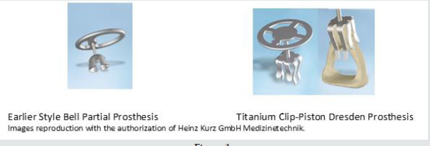
Ossicular chain reconstruction remains the primary method of restoring conductive hearing deficits commonly seen in chronic middle ear disease [2,3]. Since 1994, the German company Kurz has developed different forms of titanium ossicular prostheses. The earlier style Bell prosthesis (Bell) was designed as a partial ossicular replacement prosthesis (PORP) to be placed on the intact stapes capitulum (Figure 1). Design developments led to the newer Clip-Piston Dresden Prosthesis (Clip) which features a springloaded fastening mechanism to secure the coupling to a mobile stapes (Figure 2). The design is reported to reduce dislocation and extrusion of prosthesis with superior audiological outcomes. The purpose of this study is to compare postoperative audiological outcomes and complications of the Clip prosthesis with the Bell prosthesis.

Figure 1.

Lupinepublishers-openaccess-otolaryngology-journal

Figure 2: Age histogram.

Lupinepublishers-openaccess-otolaryngology-journal

Materials and Methods

Following institutional review board approval (no. 004834UNSW), A de-personalized chart review of all patients who underwent partial ossicular replacement using a Kurz titanium prosthesis from January 2014 to December 2018 was undertaken. All operation was performed by a single surgeon, the senior author (TRK). All the patients had cartilage interposition grafts between the prosthesis and the tympanic membrane. Cohorts were stratified based on the use of either the Kurz Clip-Piston Dresden or the oldstyle Bell Prosthesis. All procedures were analyzed in a postsurgical data base. Pre-operative and post-operative audiograms recorded thresholds at 0.5, 1, 2 and 3 KHz according to AAO-HNS guidelines. All patients completed audiograms 1 week before and three months post-surgery. These results were compared and used for statistical analysis using JASP Statistics Software (Sir Harold Jeffreys’ Statistics Program, University of Amsterdam). Successful reconstruction was determined by a post-operative air-bone gap of 20dB or less [1,4]. A Paired Sample T-test with a Wilcoxon signed rank correction was done for statistical validity.

Results and Analysis

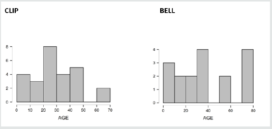
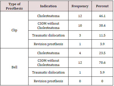
Forty-three patients underwent partial ossicular chain reconstruction during the study period. This cohort comprised both adults and children with ages ranging from 7 to 77 years (Figure 1). Of these 8 were children and 35 were adults. Follow up ranged from 6 months to 3 years with a mean of 22 months. The indications for an Ossicular Chain Reconstruction are summarized in Table 1. The most common indication overall was cholesteatoma followed closely by chronic suppurative otitis media (CSOM) without cholesteatoma.

Table 1: Indication for ossicular chain reconstruction.

Lupinepublishers-openaccess-otolaryngology-journal

Overall hearing results

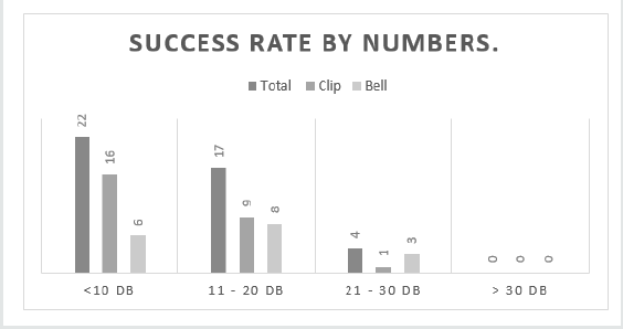
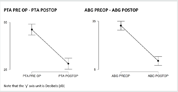
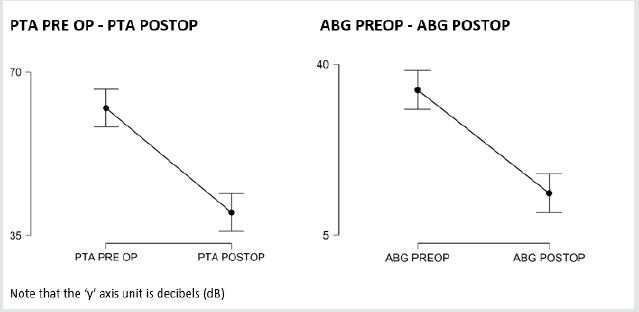
The preoperative and the postoperative audiogram are included in our statistical analyses. The pure tone audiogram (PTA) and the air-bone gap (ABG) included 0.5, 1, 2 and 3 kHz [1,5-7]. The overall hearing thresholds for the PTA and ABG values are shown in Table 2. The overall PTA improved by 20 dB +/- 0.63 (preop: 52.6 +/- 19, postop: 32.6 +/- 14.8), which is statistically significant. The overall ABG improvement was 33.9 +/- 11.4 (preop: 13 +/- 7.6, postop: 20.7 +/- 3.8). There was a very small difference in improvement between the results in Clip and Bell group for PTA (21.6 dB +/- 4.1 vs 22.3 dB +/- 4). A small difference between the results in Clip and Bell groups for ABG (21.8 dB +/- vs 21.2 +/- 0.4 dB) was also noted. Overall, A successful postoperative ABG of <20 dB was obtained in 91% of the patients (n=39) (Table 3 and Figure 2). The Clip prosthesis had a significantly higher success rate of 96% (n=26) in comparison to the Bell prosthesis which was successful in 82% of patients (n=14) (p< 0.001). Furthermore, closure of the ABG to <10dB was achieved in 61% of patients implanted with the Clip prosthesis in contrast to only 36% of those with the Bell prosthesis. A plot diagram of the paired sample T-Test comparing PTA pre and post-surgery and ABG pre and post-surgery comparing Clip and Bell prostheses show that although both prosthesis succeed in improving PTA and ABG post-operatively, the Air Bone Gap is slightly more reduced using the Clip-Piston prosthesis (Figures 3 &4). Repeated measures Anova with a Post Hoc test was used in order to compare the two prostheses. The difference between the two prosthesis was statistically significant (p <0.01). The Clip prosthesis showed a moderate effect size (Cohen’s d= -0.42) when comparing the PTA results (Table 5) and a small effect size (Cohen’s d= -0.25) in comparison of the ABG (Table 6).

Figure 3: Hearing results by Procedure.

Lupinepublishers-openaccess-otolaryngology-journal

Figure 4: Plots of PTA pre-surgery and post-surgery and ABG pre- and post-surgery using CLIP.

Lupinepublishers-openaccess-otolaryngology-journal

Figure 5: Plots of PTA pre- and post-surgery and ABG pre- and post-surgery using BELL.

Lupinepublishers-openaccess-otolaryngology-journal

Table 2: Mean overall hearing results.

Lupinepublishers-openaccess-otolaryngology-journal

Table 3: Success rates: Defined in ABG <20 dB according to AA0-HNS Guidelines.

Lupinepublishers-openaccess-otolaryngology-journal

Table 4: Post Hoc Comparisons - Type of Prosthesis PTA.

Lupinepublishers-openaccess-otolaryngology-journal

Table 5:Post Hoc Comparisons - Type of Prosthesis ABG.

Lupinepublishers-openaccess-otolaryngology-journal

Complications

One patient in the Clip prosthesis cohort required a revision procedure due to the inadequate length of the prosthesis. This was replaced by a longer prosthesis without complication. No patients experienced extrusion of their prostheses or sensorineural hearing loss. One revision OCR in the Bell cohort was needed as it extruded, subsequently it was replaced with a Clip prosthesis. No patients experienced sensorineural hearing loss.

Discussion

The titanium Clip has been designed to improve operative and hearing outcomes over previous generation of prostheses developed for ossicular chain reconstruction [8]. With flexible feet at the base of the prosthesis, a secure coupling can be achieved with the stapes capitulum with ease of application leading to reduced displacement and improved sound conduction [9-11]. This study aimed to evaluate the audiological outcomes and safety profile of this prosthesis and compare this to the earlier Bell prosthesis.
Our overall results demonstrate a significant improvement in hearing outcomes with both prostheses. The average improvements in the ABG for the Clip and Bell prosthesis were 21.8dB and 21.2dB, respectively. Similarly, favorable outcomes have also been reported in the literature. In their study of 130 patients receiving the Clip prosthesis, Kahue et al. [12] observed a reduction in the median ABG and PTA of 11dB in comparison to preoperative values. Similarly, Gostian et al. [13] demonstrated stable hearing outcomes in their long-term study (6.5 years) where a reduction in ABG of 8.9dB was seen. In our cohort of patients receiving using the Clip prosthesis, successful ossicular reconstruction, defined as an ABG of 20dB or less, was achieved in 91% of patients. This was a significant improvement over the Bell prosthesis which exhibited a success rate of 82%. Furthermore, an ABG of 10dB or less was achieved in 61% of patients in the Clip versus just 36% of those in the Bell cohort. The superiority of these outcomes is substantiated by the previously mentioned authors where success rates of 72% [13] and 63% [12] were achieved using the Clips prosthesis.
We that found that one patient experienced extrusion of the Bell prosthesis despite the use of cartilage interposition grafts. No such cases were experienced with the Clip prosthesis however one revision was required with this device due to inadequate length and persistent hearing loss. The Clip prosthesis was able to be replaced without any undue force on the stapes or subsequent complications. These low rates of displacement/extrusion compare far more favorable that those reported with earlier generation prostheses which have demonstrated displacement in up to 7.7% of cases [14]. This is likely the consequence of the clip design optimizing adherence to the stapes superstructure. The design also requires a small amount of force to engage the clip onto the stapes capitulum. In our study, this did not result in disruption of the stapes or sensorineural hearing loss in any of our patients which supports the safety profile of its application. Indeed, in the two compared studies no significant events were reported as a result of the surgery and a minor extrusion rate of 1.5% in patients which was seen in the setting of recurrent middle ear disease [12]. Several limitations can be described in this study. The retrospective nature of this investigation lends itself to inherent selection bias. This in addition to the small cohort size may influence the significance of the statistical analysis. With further recruitment and longer term follow up more definitive conclusions can be drawn and provide opportunity for subgroup analysis.

Conclusion

Partial ossicular chain reconstruction using the titanium Clip prosthesis provides excellent audiological outcomes superior to those seen with the earlier generation Bell prosthesis. Successful hearing restoration was seen in 91% of patients with a favorable safety profile. No cases of displacement, extrusion or sensorineural hearing loss were encountered.

Read More Lupine Publishers Otolaryngology Journal Articles:
https://lupine-publishers-otolaryngology.blogspot.com/


Friday, May 28, 2021

Lupine Publishers | The Discrepancy Between the Length of the Styloid Process and the Symptoms of Eagle’s Syndrome: A Case Report

 Lupine Publishers | Journal of Otolaryngology


Abstract

Eagle’s syndrome was a rare condition, and it was not commonly suspected in clinical practice. The elongation of the styloid process (SP) was considered as the main cause of this syndrome. However, many patients who were incidentally found of an elongated SP were asymptomatic. This case report presented a rare case of the bilateral elongated SP with the unilateral symptom. A 59-year-old woman who had come to our attention with the complaint of pain in the right side of the neck, and intensified pain during neck rotation, swallowing and mouth opening. She also complained of pain in the angle of the mandible, the face and otalgia. The computed tomography scans and 3D reconstruction allow us to measure the angulation and length of the SP as well as evaluate the relationship between the SP and adjacent anatomical structures. Surgical excision was performed on the right side although the patient was diagnosed with bilateral elongated SP. The postoperative course passed regularly, and the postsurgical control showed no complaint.

Keywords: Eagle’s Syndrome; elongated styloid process; stylohyoid complex

Introduction

The styloid process (SP) is a projection of thin, cylindrical and along bone from the temporal bone, and its location is between the internal and external carotid artery. The mastoid process and the tonsillar fossa are at the posterior and inside it, respectively [1]. The styloid process is one part of the stylohyoid complex including stylohyoid ligament, lesser horn of hyoid bone [2]. Initially, elongated SP syndrome or Eagle’s syndrome was described by an otorhinolaryngologists, Dr. Watt Eagle [3]. The symptoms have diversely presented with ipsilateral cervicofacial pain, referred otalgia, sore throat, dysphasia, headache, and a foreign body sensation in the pharynx. The pain regularly limits in the angle of the mandible, and neck mobility will be reduced when the head rotates to the affected side [1,4]. A physical examination is induced by digital palpation of elongated SP through the tonsillar fossa, and once palpated, the symptoms may intensify. The diagnosis is often misleading because of the vagueness of symptoms as well as the infrequent clinical observation, and these patients seek a variety of treatments in several different clinics such as dentistry, neurosurgery, neurology, psychiatry. These treatments do not relieve the symptoms, and they make the whole clinical picture cloud [4,5]. The mean length of the styloid process ranged from 20 to 25 mm [6,7]. Generally, the SP is considered elongation when it is beyond 30 mm [6,8]. There are two types of this syndrome: the classic and the carotid artery type. The former type, also known as stylalgia, always following tonsillectomy, and usually related to the elongated SP. The latter type is characterized by nonspecific symptoms that are caused by compression of the sympathetic fibers and carotid arteries, and the most common etiology of the syndrome that is the mineralization of the stylohyoid ligament [7,9]. Eagle’s syndrome is caused by an elongated SP, on the contrary, the presence of an abnormal length of SP does not result in Eagle syndrome. Here, we present a case with unilateral Eagle syndrome and bilateral elongation of the SP

Case Report

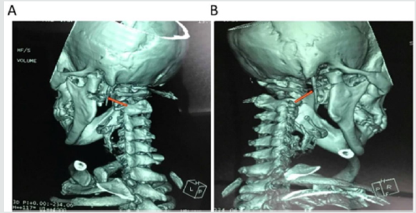
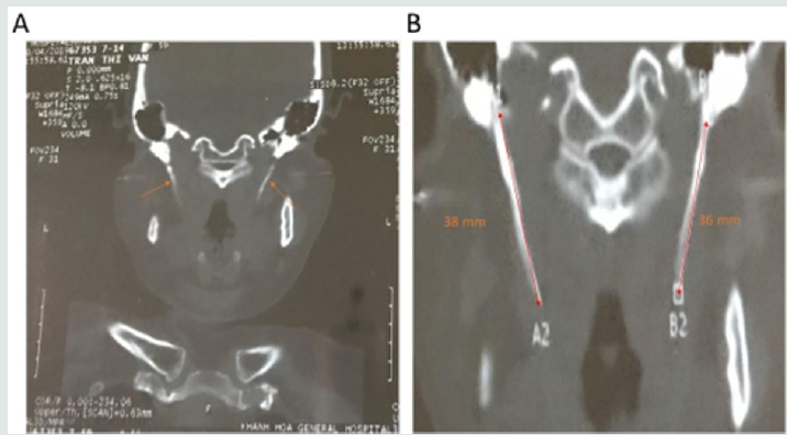
A 59-year-old woman presented to the Department of Otorhinolaryngology, Khanh Hoa general hospital, Viet Nam with the complaint of pain in the right side of the neck, the angle of the mandible, the face and otalgia that had started approximately one month previously. She simultaneously complained of intensified pain during neck rotation to the right side, swallowing and mouth opening. The patient was uneventful for any surgical or trauma history. In the physical examination, the pain was felt when palpation was performed at the angle of mandible, sternomastoid muscles. Intraoral examination, the SP was not felt on palpation of the tonsillar fossa. No particular abnormalities were detected in the video-laryngoscopic examination and neck ultrasound. Finally, the symptoms did not improve following medical therapy. Thus, the patient underwent a CT scan with the 3D reconstruction of the head and neck (Figure 1). The CT examination revealed a bilateral elongation of the SPs. The SPs were measured 36 mm on the right side and 38 mm on the left side (Figure 2). Lidocaine (2%) was deeply infiltrated into the lateral tonsillar fossa on the right side. After infiltration, immediate relief of the pain partially supported the diagnosis of Eagle’s syndrome. Although the SP on the left side was longer than another on the right side, we decided to do a right styloidectomy because of no symptom revealing on the left side. The styloidectomy was made via the intraoral surgical approach (Figure 3). Antibiotic was administered preoperatively. The surgery was under general anesthesia and the postoperative period passed regularly. The patient was discharged on the second postoperative day. At regular postoperative examinations, complete remission of symptoms was accomplished.

Figure 1: Computed tomography scan with 3D reconstruction on the left (A) and right (B) side that showed an elongation of the SPs (orange arrows).
Lupinepublishers-openaccess-otolaryngology-journal

Figure 2: A 59-year-old woman reported neck pain on right side of the face during neck rotation to right side and while swallowing and opening her mouth. CT examination showed the bilateral elongation of the SPs (A), which was long elongated on the left than on the right side (B). The SPs measured 38 and 36 mm on the left and right side, respectively (B).
Lupinepublishers-openaccess-otolaryngology-journal

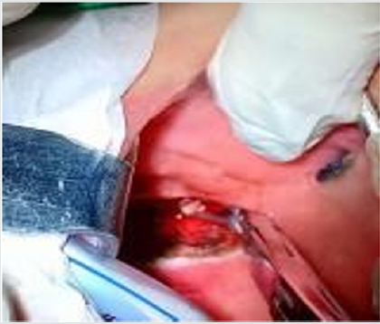
Figure 3: Intraoral approach to the styloid process. Tonsillectomy was performed first on the right side. The palpation of the tonsillar bed was performed, and the tip of the right SP was identified. The mucosa was dissected longitudinally at the point of the felt tip in the tonsillar fossa. To avoid vascular injury, the parapharyngeal space was carefully dissected by q-tips. Palpation was occasionally performed during surgery to identify the location of the SP. After the SP was exposure and excised, the tonsillar bed was carefully sutured with absorbable sutures.
Lupinepublishers-openaccess-otolaryngology-journal

Discussion

The stylohyoid complex (SC) was formed by the SP, stylohyoid ligament, lesser cornea of the hyoid and superior portion of the hyoid corpus. Embryologically, these have been derived from Reicher’s cartilage (the second branchial arch) [6,10]. Based on the successive development of the SC, it could be divided into four sections. The most proximal SC was known as the tympanohyaland which gave rise to the tympanic portion of the SP. The second portion, known as the stylohyal, forming the distal portion of the SP. The third portion (the ceratohyal) degenerated in utero, and it gave rise to the stylohyoid ligament. The most distal portion, known as the hypohyal, forming the lesser cornu of the hyoid bone [10]. The SP originated from the temporal bone behind the mastoid, and it ran anteromedially. Its anatomical variation was rarely changed in course, and it passed between the external and internal branches of the carotid artery. Cranial nerves including n. accessory, n. hypoglossus, n. vagus and n. glossopharingeus were placed medially to the SP. Three muscles (stylopharyngeus, stylohyoid and styloglossus) and two ligaments (stylohyoid and stylomandibular) were attached on the SP [2,5,10]. The length of the SP was individually variable, and the SP was considered as the elongation whenever it was longer than 30 mm. However, the existence of an elongated SP was not pathognomonic for Eagle’s syndrome, because many patients who were found of an elongated SP were asymptomatic [10]. Moreover, the elongation of the SP occurred in approximately 4% of the population [11], and only 4% of this group complained of symptoms [1,12]. Several pathophysiological mechanisms were used to explain the symptoms of Eagle’s symptoms:

a) The proliferation of granulation tissue after traumatic fracture of the SP induced the pressure on the surrounding structure [13,14].

b) Compression of adjacent nerves such as the glossopharyngeal nerve, the trigeminal nerve and the chorda tympani nerve [1].
c) Insertion tendonitis was known as a degenerative and inflammatory change which occurred in the tendinous portion of the connected area of the stylohyoid ligament [1,14].
d) The formation of granular tissue after tonsillectomy or the direct compression resulted in the irritation of the pharyngeal mucosa (involvement of the 5, 8, 9, and 10 cranial nerves) [1].
e) The impingement of the sympathetic nerve in the arterial sheath [15].

Treatment of Eagle’s syndrome was both conservative and surgical. The conservative treatment included nonsteroidal or steroidal anti-inflammatory drugs, antidepressants, anticonvulsants and exercises for the neck [1,7]. The surgical method involved amputating or removing the elongated SP via the intraoral or extraoral approach. Both approaches have been known to have pros and cons in use. The intraoral technique was simpler and took less time as well as avoided the surgical scar; however, its disadvantages were injury of the blood vessels, infection of deep neck spaces and poor visualization of the surgical field. On the other hand, the extraoral technique through cervical incision allowed better visualization of the operative field. This technique, nevertheless, took a longer time, and it could cause injury of the facial nerve. Moreover, the patient postoperatively recovery was longer and resulted in a visible scar [16-18]. Routinely, we took the intraoral approach to our patients. Due to being familiar with the technique, we have not encountered any of the complications which were mentioned above. Also, the injury of vascular and neural tissues was minimal by this method.

Conclusion

The case verifies the possibility that unilateral symptoms can occur in the bilateral elongation of the styloid process. To avoid excessive resection, the side of the styloid process should be selected by the accurate history of the patient, the tonsillar palpation and radiologic confirmation.

Conflicts of interest

Authors have none to declare

Acknowledgment

This research was supported by Basic Science Research Program through the National Research Foundation of Korea (NRF) funded by the Ministry of Education (2018R1A6A1A03025523).

Read More Lupine Publishers Otolaryngology Journal Articles:
https://lupine-publishers-otolaryngology.blogspot.com/


Choanal Atresia Repair, A Comparison Between Transnasal Puncture With Dilatation And Stentless Endoscopic Transnasal Drilling

  Abstract Background: in this study we present the outcome of surgical repair of choanal atresia of 33 patients underwent t...