Today is the time to be thankful, remember good times, and embrace those who enrich our lives. I’m thankful for a lot of things. Happy Thanksgiving to all! from our Scholarly Journal of Otolaryngology (SJO)
Today is the time to be thankful, remember good times, and embrace those who enrich our lives. I’m thankful for a lot of things. Happy Thanksgiving to all! from our Scholarly Journal of Otolaryngology (SJO)
Lupine Publishers | Journal of Otolaryngology
Dermoid cysts of the auricle are extremely rare. A 13-year-old female patient was admitted to our clinic with the complaint of a painful, slowly growing mass that had been present behind her right ear since birth. Ear examination revealed a soft, approximately 2x2 cm cystic mass on the posterior aspect of the right auricle. Histopathological examination of the excised mass was reported as dermoid cyst. The patient, who had no problem after the operation, was called to the controls and discharged. We present this case because dermoid cysts of the auricle are extremely rare and should be considered in the differential diagnosis of congenital masses in children.
Dermoid cysts are present at birth and predominantly occur in men. They are asymptomatic, slow growing, single cavity cystic masses. Most are in ovaries [1]. Less than 7% appears in the head and neck region [2].The most common location in the head and neck region is front orbital, in the upper outer part of the orbital. Other settlements are the midline of the nose,the neck, the sublingual region, and the sternal, perineal, scrotal, and sacral regions [3]. Dermoid cysts trapped in the ectoderm sac next to normal folds or surface is a developmental disorder caused by the failure of the ectoderm to leave the neural tube[4].The most valid theory about the dermoid cysts was proposed by New and Erich that was the persistence of the germ layers at birth, along the embryonic fusion line in the deep tissues in the neck. The irregular growth and differentiation of these cells causes the appearance of dermoid cysts. Dermoid cysts are divided into 3 histological types: epidermoid, dermoid and teratoma. Epidermoid cysts contain laminated keratin materials and does not contain a sebaceous gland around. Dermoid cysts are surrounded with a stratified squamous cell epithelium and they are subcutaneous tumors, and they contain various types of skin supplements such as hair follicle, sebaceous gland and sweat glands. Teratomas originate from totipotent cells, contain all three embryonic germ layers and are true neoplasms [4].
Thirteen-year-old female patient with a mass complaint in the right auricle admitted to the Otorhinolaryngology outpatient clinic of Kadirli State Hospital. The patient’s mother told that the mass existed since the birth of the child. She stated that the mass was small and painless at the beginning. For the past few years, the mass was growing and became painful. In the otorhinolaryngologic examination of the patient, there was a soft, painless, cystic mass in the posterior part of the upper inner quadrant of the right auricle just lateral to the sulcus. The mass was about 2x2 cm in size. The mass is totally excised under local anesthesia. In the macroscopic examination of specimen, the cyst was surrounded by a stratified squamous cell epithelium and the hair follicles were seen in the lumen. In the 4x10 microscopic examination with hematoxylin-eosinophil stain the cyst was surrounded by the stratified squamous cell epithelium and sebaceous glands were present in the dermis and hair follicle structures were seen in the cyst lumen. Histopathological examination results were reported as dermoid cyst.
When we look at the literature, dermoid cysts of the auricle
are extremely rare. Ikeda reported 2 cases of the dermoid cyst of the
auricle.1 Later, Samper, Bauer, Meagher and DeSouza reported
cases of postauricular dermoid cyst[4-7].In 2014, Horikiri et al.
reported dermoid cyst of the auricle [8]. Jung et al. reported a
case of the congenital dermoid cyst in the right auriculocephalic
sulcus[9]. Byeon et al. reported a dermoid cyst on the posterior of
the auricle[10]. Also,Wisevarver et al. reported a case of a dermoid
cyst on the posterior of the right auricle [11]Nasirmohtaram et al.
reported a case of a dermoid cyst located in the concha [12]. Kim
et al. reported a case of acquired dermoid cyst and stated that it is
not different from the congenital dermoid cyst. Congenital dermoid
cyst is surrounded by normal tissues the acquired dermoid cyst is
surrounded by fibrous scar tissue[13]. The differential diagnosis
of the post auricular cyst includes the epidermal inclusion cyst,
the trichilemmal cyst, the lipoma, and the hemangiomas. The
trichilemmal or the sebaceous cysts are clinically similar to the
epidermoid cysts. The diagnosis was confirmed histologically by
the presence of the amorphous keratin material in the cyst cavity.
The lipomas are common benign soft tissue adipose tumors and are
similar to the dermoid cysts. Hemangiomas are present at birth and
are benign tumors of the vascular endothelium and spontaneous
involution is possible[8]. The treatment of the dermoid cyst is
removal of the cyst wall with complete surgical excision. If it is not
removed, it may result in relapse or infection[10].The treatment
prevents the conversion to malignancy.
As a result, the dermoid cysts are rare, usually benign, and
very rarely malignant. They are congenital masses that can show
transformation and can be seen in many parts of the body. They
must be considered in the differential diagnosis of congenital ear
masses, especially in the children.
Read More Lupine Publishers Otolaryngology
Journal
Articles:
https://lupine-publishers-otolaryngology.blogspot.com/
Lupine Publishers | Journal of Otolaryngology
CHistory does not repeat itself. Though every single historical
moment is distinct, parallels can be drawn between different historical
events. Even though history does not teach us what to do, it can inspire
us to act. Revising the 1918 influenza pandemic is an opportunity to
consider the current coronavirus (COVID-19) crisis from a different
perspective. Influenza and coronavirus share basic similarities in the
way they are transmitted via respiratory droplets and contact surfaces.
Descriptions of H1N1 influenza patients in 1918-19 resemble the
respiratory failure of COVID-19 sufferers a century later. Current
discussions about holding back social distancing measures and opening
the country frequently refer to “waves” of disease that characterized
the dramatic mortality of H1N1 influenza in three major peaks in
1918-19. As COVID-19 rates begin to stabilize in some parts of the U.S.,
people today are nervously eyeing the “second wave” of influenza that
came in autumn 1918, that pandemic’s deadliest period.The 1918 influenza
pandemic took place during the First World War with three successive
waves: the first in the spring of 1918, the second – and most lethal,
responsible for 90% of deaths – in the autumn of 1918, and a final one
from the winter of 1918 to the spring of 1919. By the end of it, more
than half of the world’s population had been infected. Estimations on
mortality, showed a broad spectrum ranging from 2.5 to 5% of the world’s
population, which translates to between 50 and 100 million deaths. The
pandemic was, therefore, five to ten times deadlier than the First World
War.
Waves evoke predictability, however, and COVID-19 has been hard to
predict. Despite the lessons drawn from past influenza outbreaks, how
pandemic influenza struck in 1918 is not an exact template for what can
happen with COVID-19 in the upcoming months [1].The 2020 coronavirus and
1918 Spanish influenza pandemics share many similarities, but they also
diverge on some points. Here we empathize some of those points.
According
to Deutsche Bank, a major difference between Spanish flu and COVID-19 is
the age distribution of fatalities. For COVID-19, the elderly has been
hit the worst. For the Spanish flu of 1918, the younger population were
severely affected. The death rate from pneumonia and influenza that year
among the middle-aged population in the United States was more than 50%
higher than that for the older population. Back to COVID-19, the
overall mortality rate measured by weekly new deaths and weekly new
cases is around one-third of the level observed in the second half of
April which shows a decline in the current wave [2].Over 500 million
people, or one-third of the world’s population, became infected with the
1918 Spanish flu. According to the Centers for Disease Control and
Prevention, approximately 50 million people died worldwide, with about
675,000 deaths occurring in the US. They added that during the pandemic,
mortality was high in three categories of people: younger than 5 years
old, 20-40 years old, and 65 years and older. The high mortality in
healthy people, including those in the 20-40-year age group, was a
unique feature of this pandemic. With no vaccine to protect against it
and no antibiotics to treat secondary bacterial infections that can be
associated with it, controlling the disease worldwide were limited to
non-pharmaceutical interventions.COVID-19, the disease caused by the
virus SARS-CoV-2, has already proved extremely infectious. According to
Johns Hopkins University’s Center for Systems Science and Engineering,
it had approximately infected 13.1 million people globally and more than
3.4 million in the U.S. The disease had killed at least 573,664 lives
worldwide and 135,615 in the U.S.As for Symptomatology, for both
COVID-19 and flu, one day or more can pass between a person becoming
infected and when he or she starts to experience illness symptoms.
However, if a person has COVID-19, it usually takes longer to develop
symptoms than if they had flu. For the flu, a person develops symptoms
anywhere from 1 to 4 days after infection but for COVID-19, symptoms can
appear as early as 2 days after infection or as late as 14 days after
infection,
and the time range can vary [3].
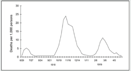
Being firstly identified in the Chinese city of Wuhan, some have labeled COVID-19 the ‘Chinese virus’. Stigmatizing a group or a nation for its alleged responsibility in a calamity is not a new trend. Take the misnomer of the ‘Spanish Flu’: unlike most of the countries at war at the time, where censorship was extreme and newspapers were initially not allowed to report on the disease, the Spanish press firstly covered the spread of the virus, creating false assumptions that the epidemic originated in Spain.Many other nicknames were given to the pandemic based on nationality or race, for example: ‘Spanish Lady’, ‘French Flu’, ‘Naples Soldier’, ‘War Plague’, ‘Black Man’s Disease’, ‘German Plague’, or even the ‘Turco-Germanic bacterium criminal enterprise’.War censorship and propaganda also had adverse effects on efforts to mitigate the pandemic. By attempting to censor information on the seriousness of the situation, many belligerent countries most certainly hindered public health efforts to stem the pandemic. Many people did not understand how the flu, an ordinarily mild illness, could cause so many deaths. Some believed their government was lying and trying to hide the return of typhus, cholera, or a so-called ‘pneumonic plague’. In Germany, some people accused the government of using a fake pathogen as a pretext to hide the deaths that were caused by malnutrition and exhaustion according to them.The lives lost during that old pandemic teach us that transparent information is essential at all times(Figure 1). To follow public health measures, the population needs to trust the authorities. In 1918, after four years of conflict and propaganda, that trust was broken. This is even more true in 2020. Mistrust of information from health authorities is still a challenge. Modern means of communication and the digital social networks make it even harder. Undocumented claims, false information, conspiracy theories, and dangerous conclusions can spread as quickly as viruses [4,5].
Figure 1: Three waves of death during the pandemic: weekly combined influenza and pneumonia mortality, United Kingdom, 1918–1919. The waves were broadly the same globally[5].

None declared.
Read More Lupine Publishers Otolaryngology
Journal
Articles:
https://lupine-publishers-otolaryngology.blogspot.com/
Abstract Background: in this study we present the outcome of surgical repair of choanal atresia of 33 patients underwent t...