Lupine Publishers | Journal of Otolaryngology
Abstract
The present case report describes the surgical removal of mucocele in a young female patient. A thirteen year old female patient was diagnosed clinically with mucocele of lower lip. A conventional surgical procedure was performed using scalpel and the excised lesion was sent for histological investigation. Histological investigation revealed well circumscribed cystic spaces filled with mucous clearly illustrating extravasation type mucocele. Complete regression of the lesion was seen with no recurrence in 6 months follow up. Surgical excision of the lesion with the involved minor salivary gland proved to give satisfactory results.
Keywords: Oral mucocele; diagnosis; excision; minor salivary gland.
Introduction
Oral mucocele are benign mucous filled cavities commonly found in oral cavity, paranasal sinuses and appendix [1,2]. It is the seventeenth most commonly found salivary lesion of the oral cavity [3]. The term mucocele was derived from Latin word (“muco”- mucous and “coele”- cavity) [4]. The characteristic features of this lesion is simple or multiple, soft, translucent and fluctuant [5]. It occurs due to accumulation of mucous due to trauma and obstruction of salivary gland duct [6,7]. There are two types of oral mucoceles generally seen in oral cavity named as, extravasation and retention type [8]. Extravasation type is most commonly found in children [9]. The most frequently affected site is lower lip and other sites include palate, buccal mucosa and floor of the mouth [10]. Although mucocele is painless, it may cause disturbances in speech and swallowing [11]. Various treatment strategies which includes surgical excision, cryosurgery, laser ablation, sclerotherapy and intralesional injection of sclerosing solutions [12]. The present study was undertaken to evaluate a case report of mucocele on lower lip treated by surgical excision using scalpel.
Case Report
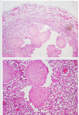
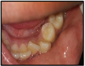
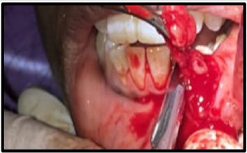
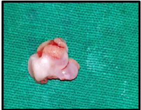
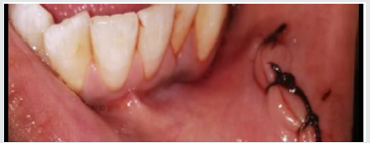
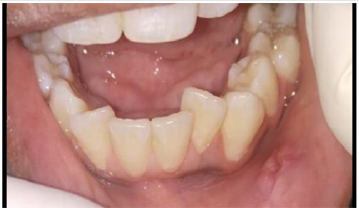
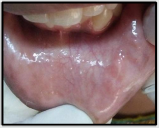
Thirteen year old female patients reported with the chief complaint of swelling on the inner aspect of her lower lip since 4 weeks. Swelling gradually increased in size and had difficulty in speech and swallowing. Patient also had a positive history of lip biting with no history of medical illness. On intra oral examination there was a small, round, fluctuant swelling seen on the labial mucosa of lower lip at the left premolar region. Swelling was 3-4 mm below the vermillion border of the lip measuring approximately 7-8 mm. Color of the swelling was similar to the adjacent mucosa (Figure 1). Based upon the clinical features and history of lip biting, the lesion was diagnosed as mucocele. The surgical procedure was carried under local anesthesia using scalpel by placing an incision circumferentially. Lesion was excised along with involved minor salivary glands (Figure 2 & 2a) and sent for histological investigations Interrupted sutures was placed (Figure 3) and removed after one week of follow up (Figure 4). Postoperative instructions were given and analgesics were prescribed. The diagnosis was confirmed as mucocele by histological report (Figure 5). Patient was followed up for 6 months and no history of recurrence was seen (Figure 6).
Histopathology
The histopathological features were suggestive of mucocele extravasation type confirming and correlating with the clinical diagnosis (Figure 5).
Figure 5: Histopathological picture (20X and 40X) showing small cystic spaces containing mucin and mucus-filled cells, areas of spilled mucin surrounded by a granulation tissue.
Discussion
Mucocele is the most common cystic swelling in the oral cavity with the incidence of 0.4 to 0.8% [13]. Traumas caused by lip biting or obstruction of duct are considered as critical factors in mucocele formation [14]. The present report illustrates a case of mucocele in lower lip which was surgically excised with the help of scalpel. The histological study revealed small cystic cavities filled with mucous enclosed by granulation tissue and inflammatory cells infiltrate. Although there are many treatment strategies for the management of mucocele conventional surgical removal is proven to be the most commonly used technique. The results were satisfactory with no signs of recurrence and minimal scar formation.
Conclusion
Although oral mucocele presents itself as an asymptomatic lesion can cause discomfort to the patient and therefore, requires treatment. Surgical excision of mucocele is a simple, cost- effective and the best alternative procedure.
Read More Lupine Publishers Otolaryngology
Journal
Articles:
https://lupine-publishers-otolaryngology.blogspot.com/

No comments:
Post a Comment