Friday, May 28, 2021

Lupine Publishers | The Discrepancy Between the Length of the Styloid Process and the Symptoms of Eagle’s Syndrome: A Case Report

 Lupine Publishers | Journal of Otolaryngology


Abstract

Eagle’s syndrome was a rare condition, and it was not commonly suspected in clinical practice. The elongation of the styloid process (SP) was considered as the main cause of this syndrome. However, many patients who were incidentally found of an elongated SP were asymptomatic. This case report presented a rare case of the bilateral elongated SP with the unilateral symptom. A 59-year-old woman who had come to our attention with the complaint of pain in the right side of the neck, and intensified pain during neck rotation, swallowing and mouth opening. She also complained of pain in the angle of the mandible, the face and otalgia. The computed tomography scans and 3D reconstruction allow us to measure the angulation and length of the SP as well as evaluate the relationship between the SP and adjacent anatomical structures. Surgical excision was performed on the right side although the patient was diagnosed with bilateral elongated SP. The postoperative course passed regularly, and the postsurgical control showed no complaint.

Keywords: Eagle’s Syndrome; elongated styloid process; stylohyoid complex

Introduction

The styloid process (SP) is a projection of thin, cylindrical and along bone from the temporal bone, and its location is between the internal and external carotid artery. The mastoid process and the tonsillar fossa are at the posterior and inside it, respectively [1]. The styloid process is one part of the stylohyoid complex including stylohyoid ligament, lesser horn of hyoid bone [2]. Initially, elongated SP syndrome or Eagle’s syndrome was described by an otorhinolaryngologists, Dr. Watt Eagle [3]. The symptoms have diversely presented with ipsilateral cervicofacial pain, referred otalgia, sore throat, dysphasia, headache, and a foreign body sensation in the pharynx. The pain regularly limits in the angle of the mandible, and neck mobility will be reduced when the head rotates to the affected side [1,4]. A physical examination is induced by digital palpation of elongated SP through the tonsillar fossa, and once palpated, the symptoms may intensify. The diagnosis is often misleading because of the vagueness of symptoms as well as the infrequent clinical observation, and these patients seek a variety of treatments in several different clinics such as dentistry, neurosurgery, neurology, psychiatry. These treatments do not relieve the symptoms, and they make the whole clinical picture cloud [4,5]. The mean length of the styloid process ranged from 20 to 25 mm [6,7]. Generally, the SP is considered elongation when it is beyond 30 mm [6,8]. There are two types of this syndrome: the classic and the carotid artery type. The former type, also known as stylalgia, always following tonsillectomy, and usually related to the elongated SP. The latter type is characterized by nonspecific symptoms that are caused by compression of the sympathetic fibers and carotid arteries, and the most common etiology of the syndrome that is the mineralization of the stylohyoid ligament [7,9]. Eagle’s syndrome is caused by an elongated SP, on the contrary, the presence of an abnormal length of SP does not result in Eagle syndrome. Here, we present a case with unilateral Eagle syndrome and bilateral elongation of the SP

Case Report

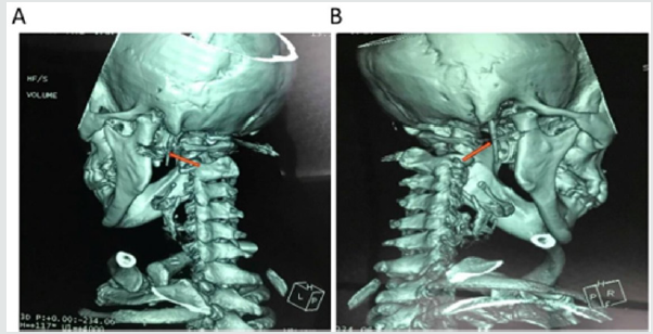
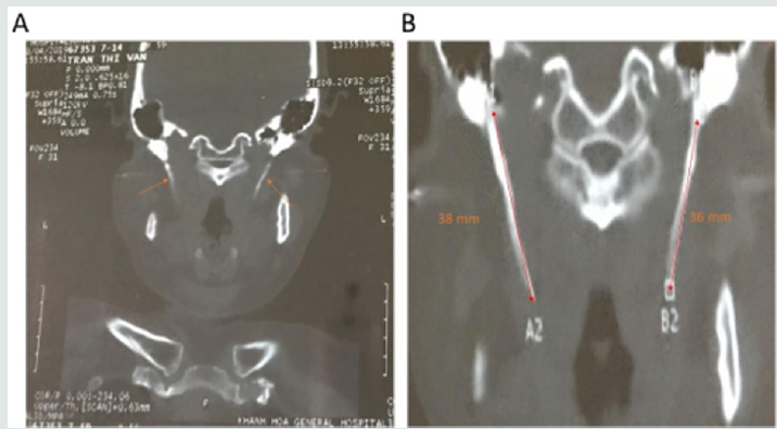
A 59-year-old woman presented to the Department of Otorhinolaryngology, Khanh Hoa general hospital, Viet Nam with the complaint of pain in the right side of the neck, the angle of the mandible, the face and otalgia that had started approximately one month previously. She simultaneously complained of intensified pain during neck rotation to the right side, swallowing and mouth opening. The patient was uneventful for any surgical or trauma history. In the physical examination, the pain was felt when palpation was performed at the angle of mandible, sternomastoid muscles. Intraoral examination, the SP was not felt on palpation of the tonsillar fossa. No particular abnormalities were detected in the video-laryngoscopic examination and neck ultrasound. Finally, the symptoms did not improve following medical therapy. Thus, the patient underwent a CT scan with the 3D reconstruction of the head and neck (Figure 1). The CT examination revealed a bilateral elongation of the SPs. The SPs were measured 36 mm on the right side and 38 mm on the left side (Figure 2). Lidocaine (2%) was deeply infiltrated into the lateral tonsillar fossa on the right side. After infiltration, immediate relief of the pain partially supported the diagnosis of Eagle’s syndrome. Although the SP on the left side was longer than another on the right side, we decided to do a right styloidectomy because of no symptom revealing on the left side. The styloidectomy was made via the intraoral surgical approach (Figure 3). Antibiotic was administered preoperatively. The surgery was under general anesthesia and the postoperative period passed regularly. The patient was discharged on the second postoperative day. At regular postoperative examinations, complete remission of symptoms was accomplished.

Figure 1: Computed tomography scan with 3D reconstruction on the left (A) and right (B) side that showed an elongation of the SPs (orange arrows).
Lupinepublishers-openaccess-otolaryngology-journal

Figure 2: A 59-year-old woman reported neck pain on right side of the face during neck rotation to right side and while swallowing and opening her mouth. CT examination showed the bilateral elongation of the SPs (A), which was long elongated on the left than on the right side (B). The SPs measured 38 and 36 mm on the left and right side, respectively (B).
Lupinepublishers-openaccess-otolaryngology-journal

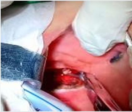
Figure 3: Intraoral approach to the styloid process. Tonsillectomy was performed first on the right side. The palpation of the tonsillar bed was performed, and the tip of the right SP was identified. The mucosa was dissected longitudinally at the point of the felt tip in the tonsillar fossa. To avoid vascular injury, the parapharyngeal space was carefully dissected by q-tips. Palpation was occasionally performed during surgery to identify the location of the SP. After the SP was exposure and excised, the tonsillar bed was carefully sutured with absorbable sutures.
Lupinepublishers-openaccess-otolaryngology-journal

Discussion

The stylohyoid complex (SC) was formed by the SP, stylohyoid ligament, lesser cornea of the hyoid and superior portion of the hyoid corpus. Embryologically, these have been derived from Reicher’s cartilage (the second branchial arch) [6,10]. Based on the successive development of the SC, it could be divided into four sections. The most proximal SC was known as the tympanohyaland which gave rise to the tympanic portion of the SP. The second portion, known as the stylohyal, forming the distal portion of the SP. The third portion (the ceratohyal) degenerated in utero, and it gave rise to the stylohyoid ligament. The most distal portion, known as the hypohyal, forming the lesser cornu of the hyoid bone [10]. The SP originated from the temporal bone behind the mastoid, and it ran anteromedially. Its anatomical variation was rarely changed in course, and it passed between the external and internal branches of the carotid artery. Cranial nerves including n. accessory, n. hypoglossus, n. vagus and n. glossopharingeus were placed medially to the SP. Three muscles (stylopharyngeus, stylohyoid and styloglossus) and two ligaments (stylohyoid and stylomandibular) were attached on the SP [2,5,10]. The length of the SP was individually variable, and the SP was considered as the elongation whenever it was longer than 30 mm. However, the existence of an elongated SP was not pathognomonic for Eagle’s syndrome, because many patients who were found of an elongated SP were asymptomatic [10]. Moreover, the elongation of the SP occurred in approximately 4% of the population [11], and only 4% of this group complained of symptoms [1,12]. Several pathophysiological mechanisms were used to explain the symptoms of Eagle’s symptoms:

a) The proliferation of granulation tissue after traumatic fracture of the SP induced the pressure on the surrounding structure [13,14].

b) Compression of adjacent nerves such as the glossopharyngeal nerve, the trigeminal nerve and the chorda tympani nerve [1].
c) Insertion tendonitis was known as a degenerative and inflammatory change which occurred in the tendinous portion of the connected area of the stylohyoid ligament [1,14].
d) The formation of granular tissue after tonsillectomy or the direct compression resulted in the irritation of the pharyngeal mucosa (involvement of the 5, 8, 9, and 10 cranial nerves) [1].
e) The impingement of the sympathetic nerve in the arterial sheath [15].

Treatment of Eagle’s syndrome was both conservative and surgical. The conservative treatment included nonsteroidal or steroidal anti-inflammatory drugs, antidepressants, anticonvulsants and exercises for the neck [1,7]. The surgical method involved amputating or removing the elongated SP via the intraoral or extraoral approach. Both approaches have been known to have pros and cons in use. The intraoral technique was simpler and took less time as well as avoided the surgical scar; however, its disadvantages were injury of the blood vessels, infection of deep neck spaces and poor visualization of the surgical field. On the other hand, the extraoral technique through cervical incision allowed better visualization of the operative field. This technique, nevertheless, took a longer time, and it could cause injury of the facial nerve. Moreover, the patient postoperatively recovery was longer and resulted in a visible scar [16-18]. Routinely, we took the intraoral approach to our patients. Due to being familiar with the technique, we have not encountered any of the complications which were mentioned above. Also, the injury of vascular and neural tissues was minimal by this method.

Conclusion

The case verifies the possibility that unilateral symptoms can occur in the bilateral elongation of the styloid process. To avoid excessive resection, the side of the styloid process should be selected by the accurate history of the patient, the tonsillar palpation and radiologic confirmation.

Conflicts of interest

Authors have none to declare

Acknowledgment

This research was supported by Basic Science Research Program through the National Research Foundation of Korea (NRF) funded by the Ministry of Education (2018R1A6A1A03025523).

Read More Lupine Publishers Otolaryngology Journal Articles:
https://lupine-publishers-otolaryngology.blogspot.com/


Saturday, May 22, 2021

Lupine Publishers | How to Prevent Nasal Obstruction After Rhino Septoplasty. Could A Routine Turbinate Reduction Improve the Outcomes in Quality of Life of Patients?

 Lupine Publishers | Journal of Otolaryngology


Abstract

The Objective of this article is to Review current literature about performing a turbinectomy associated with Rhinoseptoplasty. Three clinical trials with level one of evidence about the issue have been published recently. All of them selected patients with nasal obstruction who were submitted to Rhinoseptoplasty. The NOSE scale to measure quality of life in these patients was used. Other tools of objective measurement as Acoustic rhinometry and rhinomanometry or subjective scales, Snot-20 WHOQOl and ROE were also explored. Each study used a different technique for turbinate reduction. All of the three found the same results discussed below. To review the scientific evidences of these articles can bring new outlook about this controversial topic.

Abbreviations: QOL: Quality of Life; NOSE: Nasal Obstruction Symptom Evaluation; ROE: Rhinoplasty Outcome Evaluation

Mini- Review

Rhinoplasty is often performed to restore nasal function and form. The development or maintenance of nasal obstruction after rhinoplasty is a complication that negatively affects quality of life (QOL), and priority should be given to prevention strategies [1]. However, the available surgical techniques to prevent this obstruction have been empirically developed and are often used based on the surgeon’s preference rather than on objective criteria. Currently, strategies like spreaders grafts, support grafts, reconstruction or repositioning cartilages and even a good septoplasty are used to enlarge the nasal valve [2-6]. Another technique widely used is the Reduction of the inferior turbinate [3,7-9]. Otherwise, an established technique to reduce turbinate with hypertrophy is still debatable [10-13]. Reviews pointed that research in this field appears to be driven by technological advancement rather than by establishment of patientsʹ benefit. Partly, because of the lack of properly conducted randomized controlled trial with long term results. Some articles even question the efficacy of this procedure in cases of nasal obstruction explained for other reasons rather than turbinate hypertrophy isolated [14]. A Recent clinical trial reveal that the association of turbinectomy with septoplasty, though widespread, does not improve the nasal obstruction clinical outcomes and can add risks to patients [15].
Therewithal an objective standardized tool that links anatomy measures with clinical results is not available yet [16]. To address this issue, Stewart et al. have developed and validated the Nasal Obstruction Symptom Evaluation (NOSE) scale, a disease specific QOL instrument designed to determine the presence of nasal obstruction [17]. Since then, several studies have compared preoperative versus postoperative NOSE scores to assess QOL associated with nasal obstruction. A recent survey by the American Society of Plastic Surgeons shows that 90% of surgeons address the inferior turbinate in at least a portion of their cases, with 8% routinely reducing the turbinate in all cases. However, 10% of the respondents in this survey did not address the inferior turbinate in any of their cases [18]. Such variability in addressing this potential cause of/risk factor for nasal obstruction deserves closer attention. Guyuron [19] has pointed out that the position of the inferior turbinates contributes to airway narrowing after nasal bone osteotomy. On account of that, surgical treatment of inferior turbinates seems to be a good option to avoid postoperative nasal obstruction, which would be great because of the accessibility, simple technique and relative low risks. Unfortunately, all three latest trials could not prove any improvement in QOL when the turbinate reduction is associated even by using different techniques. Furthermore, to access the turbinate does not seem to improve the rates of nasal obstruction and satisfaction with respiratory scales outcomes [20-22].
In 2013, Lavinsky-Wolff et al. [20] compared QOL in patients undergoing primary Rhinoseptoplasty, with or without turbinate reduction by submucosal electrocautery. There was no difference between subjects submitted or not to inferior turbinate reduction in NOSE score (-75% vs. -73%; P = 0.893); all WHOQOL-bref score domains (P > 0.05), NO-VAS (-88% vs. -81%; P = 0.89) and acoustic rhinometry recordings (P > 0.05). Besides the literature does not show difference between the techniques, this study receives some critique about the conservative reduction by submucosal electrocautery chosen. In order to answer this question de Moura et al. [21], in 2017, randomized other 50 patients undergoing primary Rhinoseptoplasty associated with inferior turbinate reduction through endoscopic partial inferior turbinectomy (EPIT) reduction or not. There was no difference between the groups in absolute score changes for NOSE (-50.5 vs. -47.6; P = 0.723), Rhinoplasty Outcome Evaluation (ROE) (47 vs. 44.8; P=0.742), and all (WHOQOL-bref) score domains (P >0.05) (Table 1). There were no differences between the groups regarding presence of the complications. Surgical duration was higher in the EPIT group (212 minutes ±7.8 vs. 159.1±5.6; P > 0.001). Both articles do not present any improvement at short-term outcomes (three months). Nevertheless, a long-term result was needed to reinforce these findings. Wherefore this year Sommer et al. [22] published a clinical Trial with nine months follow up. They randomized patients to perform anterior turbinoplasty or not during septo- or Rhinoseptoplasty.

Table 1: Source de Moura et al. [21].

Lupinepublishers-openaccess-otolaryngology-journal

Dependent variable Δ scores = (postoperative score-preoperative score)
*P Value: ANCOVA of Δ adjusted for baseline NOSE -p value, nasal itching, rhinorrhea and use of spreader graft.
†P Value: ANCOVA of Δ adjusted for baseline ROE score, nasal itching, rhinorrhea and use of spreader graft.
*P Value: ANCOVA of Δ adjusted for baseline WHOQOL -bref score, nasal itching, rhinorrhea and use of spreader graft.
ANCOVA: analysis of covariance; CI: Confidence Interval; EPIT: Endoscopic Partial Inferior Turbinectomy; NOSE-p: Nasal Obstruction Symptom Evaluation Portuguese; ROE: Rhinoplasty Outcome Evaluation; SD: Standard Deviation; WHOQOL-bref: World Health Organization Quality of Life Scale

The results enhanced previous trials. Patient satisfaction after functional septo- and septorhinoplasty is high and does not seem to be affected by turbinate surgery. There was no statistically significant difference in the postoperative results regarding objective rhinological measurements with or without turbinoplasty (Table 2). They concluded that extensive resections of the turbinates can have a negative impact on nasal physiology, so the indication for turbinoplasty must be carefully considered. Considering these results, clearly has no reason to proceed a turbinate reduction, at least as routine, to patients submitted at rhinoplasty. As medical science is not so hard, presumably some phenotypes of noses probably could benefit of it. Although these patients are not identified, at least it can be justifying by other reasons, this turbinate access should be avoided. This finding changes the focus of discussion to which method should be used to reduce the turbinate to there are another surgical strategy that could be used to improve our functional results and which technique is it. Be like these finds fortify positively the discussion about structured Rhinoplasty and the importance of the reconstruction and reinforce of the nasal valve.

Table 2: Changes in MCA2.

Lupinepublishers-openaccess-otolaryngology-journal

Conclusion

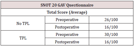
The indications for the reduction of the turbinate were well established in context of a turbinate inferior hypertrophy [23,24]. The studies have not shown, until now, a superior technique for the inferior reduction. Although techniques which preserve mucosa and have partial resection instead of total resection are indicated [10-12]. Trends in Rhinoplasty research do not show relevant benefits at patients’ quality of life outcomes associated with nasal obstruction when Rhinoplasty is performed combined with reduction turbinate. More clinical trial must be develop comparing other methods of enlargement and preservation of nasal valve and objective measurement instruments need to be developed to clarify these findings [25] (Table 3).

Table 3: Pre-and Postoperative values of the SNOT 20 GAV questionnaire within the groups (TPL vs. No TPL).

Lupinepublishers-openaccess-otolaryngology-journal

Read More Lupine Publishers Otolaryngology Journal Articles:
https://lupine-publishers-otolaryngology.blogspot.com/


Saturday, May 15, 2021

Lupine Publishers | Cancer of the Mobile Tongue: About 29 Cases Collected at the ENT and CCF Service of the University Hospital Centre of Fann (Dakar/Senegal)

 Lupine Publishers | Journal of Otolaryngology


Summary

Objectives: We report the epidemiological, clinical, paraclinical and therapeutic data of mobile tongue cancers operated in the ENT and CCF service of the University Hospital of Fann (Dakar/Senegal).

Patients and methods: This were a monocentric retrospective study, conducted at the ENT and cervico-facial surgery department of the CHNU of Fann from 1 January 2009 to 31 December 2018, i.e. 10 years. All patients operated during this period with cancer of the mobile tongue with histological confirmation were included.

Results: A total of 29 records were collected. They were 17 males and 12 females, for a sex ratio of 1.07. The mean age of the patients was 52 years with a median age of 49 years. Tobacco intoxication was found in 5 patients or 17.24% of cases. Isolated glossodynia was the most frequent symptom, (27.5%) of the cases. The lesion was ulceroburging in 55% of cases and was located on the right lateral edge of the mobile tongue in 72.4% of cases. As part of the extension workup, CT scan was performed in 4 patients. Panendoscopy was performed in 27 patients or 93.1% of cases. The tumor was classified as T3- T4 in 61.9% of cases and N0-N1 in 58.5% of cases. Hemi glossectomy was the most common procedure performed (48.27%) associated with complete unilateral functional curettage (41.3%). The anatomopathological result of the operation was in favor of squamous cell carcinoma in 100% of cases. Nine of our patients had benefited from additional treatment after surgery, i.e. 31% of cases.

Conclusion: Despite surgical advances and radiotherapy, treatment is often a failure for many at-risk patients. We conclude from this work that the prognosis is significantly better in patients diagnosed at an early stage.

Keywords: Mobile tongue; squamous cell carcinoma; glossectomy

Abbreviations: ADT: Aerodigestive Tract; SPSS: Statistical Package for Social Science

Introduction

Cancers of the tongue are the most common cancers of the oral cavity with poor survival [1,2]. The main risk factors are mainly tobacco and alcohol intoxication and poor oral health, but other carcinogens are emerging: viral (HPV) and environmental (asbestos) factors [3-5]. Like all cancers of the upper aerodigestive tract (ADT), squamous cell carcinoma is the most common histological type of cancer of the tongue in 95% of cases. It generally occurs in males from the age of 55 onwards. It is locally aggressive, and locoregional evolutionary pursuits are frequent [6-8]. Pre-therapeutic evaluation and monitoring of these cancers has been improved in recent decades by advances in medical imaging (MRI). Treatment is based on a combination of surgery and radiotherapy. Mortality from this cancer remains high: 40% survival in 5 years at all stages [2]. The aim of this study was to report the epidemiological, clinical, paraclinical and therapeutic aspects of mobile tongue cancer that has undergone surgery in the ENT and CCF department of the Fann University Hospital (Dakar/ Senegal).

Patients and Methods

This is a retrospective study conducted at the ENT and CCF service of the CHNU of Fann (Dakar/Senegal) on patients operated on for mobile tongue cancer during the period from 1 January 2009 to 31 December 2018. Excluded from this study were the records of patients with cancer of the base of the tongue, patients who were received as second hand after a first operation in another structure and incomplete records. The parameters studied were age, sex, promoting factors, clinical and paraclinical signs, type of surgery, histological result and complementary treatment. The data obtained were entered into Excel and analyzed by the statistical package for social science (SPSS) version 20.

Resultss

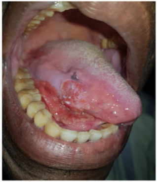
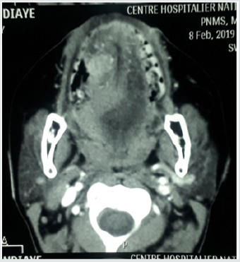
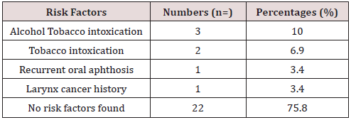
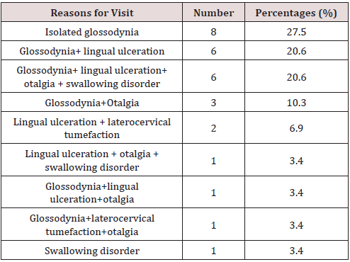
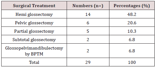
In 10 years, we have collected 29 cases of mobile tongue cancer operated on. We noted 17 male subjects, i.e. 58.6% of the cases and 12 female subjects, i.e. 41.4% of the cases with a sex ratio of 1.41. The sex ratio was 1.41. The mean age of our patients was 52 years with extremes ranging from 14 to 78 years with a median age of 49 years. Alcohol and smoking intoxication were found in 3 patients (Table 1). The average time of disease progression was 5.6 months with extremes ranging from 2 months to 12 months. Isolated glossodynia was the main reason for consultation (27.5%), (Table 2). Macroscopically, the lesion was ulcerous and budding (Figure 1) in 55% of cases, ulcer infiltrating in 17.2% of cases, ulcerous budding and infiltrating in 10.3% of cases, ulcerous budding and necrotic in 6.8% of cases, ulcerated in 3.4% of cases, infiltrating in 3.4% of cases and ulcerous budding and necrotic-infiltrating in 3.4% of cases. This lesion was located on the right lateral edge of the mobile tongue in 21 patients, i.e. 72.4% of cases. Palpation showed an extension towards the floor of the mouth in 8 patients, towards the base of the tongue without going beyond the midline in 2 patients and towards the tip of the tongue and the midline in 4 patients. The size of the lesion was estimated in 20 patients or 68.9% of cases. It averaged 3.8 cm with extremes of 2 cm to 5 cm. Palpable lymphadenopathy was found in 65.5% of cases. As part of the extension workup, a CT scan of the oral cavity (Figure 2) and cervico-thoracic cavity was performed only in 4 patients, none of the patients in our study benefited from magnetic resonance imaging. Chest X-rays were performed in all our patients. A first panendoscopy of the upper ADT was performed in 27 patients, i.e. 93.1% of the cases. Histological analysis was in favor of squamous cell carcinoma in 93.1% of cases. At the end of the extension assessment, the lesions were classified as T3-T4 in 61.9% of cases and N0-N1 in 58.5% of cases according to the TNM classification of 2017. Haemiglossectomy was the most common surgical procedure performed (48.27%) followed by pelviglossectomy (20.68%) (Table 3). Four patients had received a flap as part of tongue reconstruction. Seven patients in our study had received postoperative radiotherapy, 24.13% of the cases, and 7 other patients had received chemotherapy, of which in 2 patients it was neo-adjuvant, in 2 others adjuvant chemotherapy and in 3 patients it was palliative. The histological findings of the surgical specimen and its excision product were in favor of squamous cell carcinoma in all our patients. For the surgical specimen, the resection margins were healthy in 55% of cases and invaded in 13.79% of cases. The excision product was the site of a carcinomatous focus in 58.62% of cases and free of any lesion in 24.13% of cases. For an average delay of 9 months with extremes ranging from 1 to 32 months, we had noted 6 cases of complete remission, 8 cases of progressive continuation, no cases of recurrence and 15 patients lost sight of after the complete healing of the surgical wound.

Figure 1: Ulcer-budding lesion of the right lateral border extend to oral floor area.

Lupinepublishers-openaccess-otolaryngology-journal

Figure 2: CT scan of the oral cavity: axial section showing a malignant lesion of the mobile tongue.

Lupinepublishers-openaccess-otolaryngology-journal

Table 1: The different risk factors found in patients.

Lupinepublishers-openaccess-otolaryngology-journal

Table 2: Distribution of Patients by Reasons for Visit.

Lupinepublishers-openaccess-otolaryngology-journal

Table 3: Distribution of Patients by Surgical Treatment.

Lupinepublishers-openaccess-otolaryngology-journal

Discussion

The oral cavity is the most common location for cancers of the upper aerodigestive tract (QDT). Worldwide, cancer of the oral cavity accounts for 275,000 new cases per year, i.e. 54% of all cases of cancers of the upper aerodigestive tract, of which 40-50% occur in the tongue [1,6]. Tongue cancer represents 1.6% of all cancers in the USA [9], 1.4% in Tunisia [10] and 2% worldwide [11]. In our study the sex ratio was 1.41 in favor of the male sex. This is consistent with the results of Diombana et al. [12], Bousaadani et al. [1], Beldgadi et al. [13]. However, in some series, the sex ratio tends to shift in favour of the female sex due to the increase in the number of women who smoke from day to day [14-16]. Cancer of the tongue is a cancer of the mature subject. However, cases below the age of 30 are not exceptional, as is the case in our study with a 14-year-old patient. Bouykhelef et al. [17] report a mean age of 57 years with extremes from 34 to 80 years, EL idrissi et al. [14] report a mean age of 60 years with extremes from 36 to 80 years. In our series, we found a mean age of 52 years with extremes from 14 to 78 years. These results are close to those in the literature [13]. Considered since 1954 as a risk factor for upper ADT neoplasia, through the production of irritating substances and polycyclic aromatic hydrocarbons, tobacco intoxication is found in 60 to 80% of patients with tongue cancer [2,18,19]. Rothmann and Keller [20] specify the relationship between the intensity of tobacco consumption and the appearance of oral cavity cancer: for a risk of 1 in non-smokers, it is 1.52 if consumption is 20 cigarettes/d and 2.43 if consumption exceeds 40 cigarettes/d. Laurent [21], in a series of 70 patients with oral cancer between 1992 and 2002, found that 60% of patients were smokers with a consumption of more than one pack a day. Aksu [16] found 81% smokers, of which 38% are heavy smokers (≥ 20 cigarettes /d). The effects of smoking are potentiated by alcohol intoxication. In our study, alcohol-smoking intoxication was found in 10.3% of cases and smoking intoxication alone in 6.9% of cases. This low percentage could be explained by the lack of sincerity in the declaration of this subject or the existence of other risk factors such as viral factors (HPV) and poor oral health. The last one would probably be related to often poor socio-economic conditions, which constituted 80.5% in Bouyekhelef [17] and 58.5% in EL Idrissi [14]. However, Rakotoarison [22] in his observation found a case of squamous cell carcinoma of the tongue due to chronic irritation of a poorly adapted dental prosthesis. In our series, we reported a case of squamous cell carcinoma in a patient previously treated by radiation for laryngeal cancer. In the literature, cases of tongue cancer on precancerous lesions were reported in 5% according to [23]. These are epithelial alterations that may or may not progress to squamous cell carcinoma. They are observed mainly in men over 50 years of age and are associated with alcohol and tobacco consumption. The role of pollution and HPV are still being discussed [23]. They are mainly represented by leukoplakia and dysplasia. Cancers of the tongue are often discovered late because of the discretion of their symptomatology [2,24,25]. In our series, this delay was on average 5.6 months, which corroborates the results of El Idrissi [14]. This delay in consultation may be linked to the neglect and trivialization of symptoms by our patients or to the use of traditional medicine.

In our series, isolated glissandi were the main reason for consultation in 27.5% of cases, followed by glissandi associated with lingual ulceration in 20.6% of cases. For Belgadi [13] and Laurent [21], lingual ulceration was the main reason for consultation. In the literature the electroburning form is the most frequent form of cancer of the mobile tongue [16,17]. In our study, we found an electroburning form in 55% of cases followed by an ulcer infiltrating form in 17.2% of cases. The extension is preferentially in the anteroposterior direction but also in the thickness of the lingual muscle mass. The anterior and lateral buccal floor, the lower gum, the base of the tongue and the elements of the tonsil chamber are successively examined. In the literature, examination of the lymph node areas reveals lymph node invasion (the submaxillary, sub digastric and supra omohyoid nodes) in the order of 20% for T1, 50% for T2 and T3, and 75% for T4 [17,23]. Node involvement was found in 65.5% of the cases in our study. Brasnu et al. [26] recommend that as part of the extension assessment, MRI, PET scan, pan endoscopy, CT scan with contrast agent injection and metastatic assessment (thoracic CT scan, abdominal ultrasound and esophageal microscopy) should be performed. MRI may be offered in first line for lingual carcinoma, especially if an extension to the floor of the mouth or to the midline is suspected by palpation [2,17,18]. These different examinations could not be performed in our series due to the low socio-economic standard of living of our patients. At the end of the clinical examination and the extension assessment, EL Idrisi [14] had concluded in its work at a rate of 83% for T1 and T2 tumors. In our series, patients were classified as T3-T4 in 61.9% of cases and N0-N1 in 28.5% of cases. This result can be explained by the delay in consultation and the aggressiveness of lingual cancer. Therapeutically, the most common surgical procedure performed in our study was hemi glossectomy in 48.27% of cases and pelviglossectomy in 20.68% of cases. This is due to the fact that more than the majority of our patients were diagnosed at a late stage (61.9% T3-T4). EL Idrissi [14] performed in its series 50% partial glossectomy and 16.6% hemi glossectomy. Hicks [27] performed partial glossectomy in 74.7% followed by hemi glossectomy in 14% of cases. Post-operative radiotherapy is indicated in cases of invaded resection limits, vascular emboli, perineural invasion, lymph node involvement with capsular rupture for all tumors except T1. It is associated with concomitant platinumbased chemotherapy in the case of positive cross-examinations and extra capsular lymph node extension [4]. In our series, only seven patients received post-operative radiotherapy because at that time there was only one radiotherapy center in our country for all cancer patients.

Conclusion

Despite surgical advances and radiotherapy, treatment is often a failure for many at-risk patients. We conclude from this work that the prognosis is significantly better in patients diagnosed at an early stage.

Read More Lupine Publishers Otolaryngology Journal Articles:
https://lupine-publishers-otolaryngology.blogspot.com/


Choanal Atresia Repair, A Comparison Between Transnasal Puncture With Dilatation And Stentless Endoscopic Transnasal Drilling

  Abstract Background: in this study we present the outcome of surgical repair of choanal atresia of 33 patients underwent t...