Friday, June 25, 2021

Lupine Publishers | A Case Report of Tenosynovial Giant Cell Tumor in an Unusual Location: The Supraclavicular Region

 Lupine Publishers | Journal of Otolaryngology


Abstract

Tenosynovial giant cell tumor (TSGCT) is a benign tumor that commonly presents in the upper limb joints and hands and less frequently occurs in the lower extremities. Usually they arise from synovium or tendon sheath. The aim of this article is to present an unusual location of TSGCT of the supraclavicular region in a young patient.

Keywords: Tenosynovial, giant cell tumor, supraclavicular region

Abbreviations: TSGCT: Tenosynovial Giant Cell Tumor, CSFIR: Colony Stimulating Factor 1 Receptor, WHO: World Health Organization

Introduction

Tenosynovial giant cell tumor (TSGCT) is a benign tumor that commonly presents in the upper limb joints and hands and less frequently occurs in the lower extremities. Usually they arise from synovium or tendon sheath [1,2] .TSGCT is divided into two main subtypes: the localized type that is otherwise known as giant cell tumor of tendon sheath that is well circumscribed and occurs in small joints e.g. fingers; it has a local recurrence rate of about (4- 30%) [1,2]. The second type is the diffuse TSCGT otherwise known as pigmented villonodular synovitis that is poorly circumscribed. It occurs usually in large joints and is more aggressive than type 1 TSCGT and has a recurrence rate of 18-50%. Type 2 TSCGT is cured with radical excision [2]. In this article, we report an unusual location of TSGCT of supraclavicular region, which is the second case that is presented in the literature [3].

Case Report

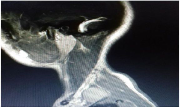
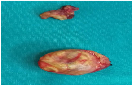
A 16-years old woman presented with a six-month history of slowly growing painless mass in left supraclavicular region. That was initially suspicious of a pathological lymph node, neurinoma or a hematological disorder (lymphoma). Neurological and vascular examination were unremarkable and laboratory values were within normal range. The patient underwent an ultrasound guided fine needle aspiration of the tumor that showed benign large multinucleated giant cells, histiocyte and xanthoma cells positive for CD68 by immunohistochemistry. The tumor showed no necrosis. The patient underwent further investigations including magnetic resonance imaging and angiography of neck (MRI and MRA scans). The results showed a 23x17x35 mm left supraclavicular mass that distorted scalene muscles in this region with normal vascular anatomy (Figure 1). The patient had an elective operation and the tumor was totally excised from level 5b left neck region and 2 lymph nodes were also removed (Figure 2). Postoperatively, the patient had an unremarkable recovery and she was discharged three days later. The histology confirmed the diagnosis of Tenosynovial giant cell tumor of left supraclavicular region. The lymph nodes showed no pathology.

Figure 1: Sagittal view MRI scan neck showing left posterior triangle mass.

Lupinepublishers-openaccess-otolaryngology-journal

Figure 2: Left supraclavicular tumor (bottom) and tissue with 2 lymph nodes(top).

Lupinepublishers-openaccess-otolaryngology-journal

Discussion

TSGCT or giant cell tumor of tendon sheath is a rare benign lesion involving joint synovia, bursae or tendon sheath. The annual incidence of TSGCT is 1.8 per million. It may be intra-articular or extra-articular and can be classified as local or diffuse lesions [1,4]. TSGCT is more common in women and the ratio is male to female 1:1.5 with average age between (30-50yrs) [1]. TSGCT may also affect children but the incidence for both localized and diffuse types is rare [5,6]. TSGCT is a monoarticular disease affecting typically large joints e.g. the knee. The second most affected joints are the hand and wrist followed by the hip and ankle. Common symptoms described in literature include pain, stiffness and swelling of the affected joint [7]. The tumor that we present is in an unusual site and according to literature is the second one described in the supraclavicular region [3]. Malignant tumors such as synovial sarcoma and benign tumors should be kept in mind as potential differential diagnosis [8,9]. Multinucleated giant cells are a major clue for diagnosis of TSGCT. On immunohistochemistry the histiocyte cells are positive for markers like CD68, CD45 and CD163 like it occurred in our case [10]. World Health Organization (WHO) classified TSGCT as a benign tumor. There is debate however whether it is a true neoplasm as studies have supported the presence of translocation in chromosome 1p11-13 and overexpression of macrophage colony stimulating factor 1 receptor (CSFIR) [7,11]. Initial treatment is surgical resection and if recurrence occurs then additional radiation therapy, synovectomy and external beam radiation therapy are recommended [12].

Conclusion

TSGCT is a benign tumor of tendon sheath of slow progression. As TSGCT in our case is located in an unusual location it is difficult to diagnose it. Diagnosis requires MRI scan and histologic confirmation. Initial treatment is surgical resection of the tumor. Recurrence rate does occur in TSGCT and therefore a close follow up is recommended.

Conflict of Interest

The authors have no conflict of interest to declare.

Read More Lupine Publishers Otolaryngology Journal Articles:
https://lupine-publishers-otolaryngology.blogspot.com/


Friday, June 18, 2021

Lupine Publishers | Transoral Robotic Resection of Lingual Thyroid

 Lupine Publishers | Journal of Otolaryngology


Introduction

Surgical removal of ectopic lingual thyroid is indicated in symptomatic patients or in cases of suspected malignancies. An external approach is most often used but is associated with morbidity and visible scar on cervical region. In this case we utilized Transoral Robotic surgery [TORS] to excise ectopic lingual thyroid tissue. Ectopic thyroid refers to the presence of thyroid tissue in locations other than the normal anterior neck region between the second and fourth tracheal cartilages. It is the most frequent form of thyroid dysgenesis, accounting for 48-61% of the cases [1]. Lingual thyroid is a rare developmental anomaly originating from aberrant embryogenesis during the passage of the thyroid gland through the neck. Prevalence of this condition is reported to be between 1 per 100,000-300,000 persons and occur one in 4,000- 8,000 patients with thyroid disease [2]. To date, about 500 cases have been reported in the English literature mainly from Europe, Asia and America, with a small number of reports coming from Africa. Sixty-five to eighty percent of cases occurred in females [2]. Although it is difficult to detect asymptomatic thyroid ectopy, postmortem studies have suggested that 7-10% of adults may harbor asymptomatic thyroid tissue along the path of the thyroglossal duct [3]. In 1869, Hickman reported the first case of ectopic thyroid tumour of the base of the tongue, pressing down the epiglottis on the larynx and causing death by suffocation sixteen hours after birth [4]. In 1999, da-Vinci robot was developed by intuitive company in USA. It contains high magnification 3-D endoscope, endo-wristed instruments with motion scaling and tremor filtration function that help surgeons to perform endoscopic surgery with more precision, dexterity and control. conventional transoral approach has the disadvantages of limited exposure and difficulty in manipulation and hemostasis [5]. In this paper we report a case of 16 years old lady from Nepal who presented to our hospital with choking symptoms which are more during night since years. She was treated as an asthmatic and had no improvement with bronchodilators. She was referred to our center for further evaluation to rule out any ENT cause. Fiberoptic laryngoscopy lead to provisional diagnosis of lingual thyroid. She underwent excision of ectopic thyroid tissue by Da Vinci robotic surgical system. Histopathological examination confirmed lingual thyroid (Figure 1).

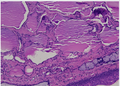
Figure 1: Photomicrograph of histology section, Hematoxylin Eosin stain, 10 X magnification, showing covering of normal squamous epithelium of tongue, with many variable sized colloids filled thyroid follicles in the subepethelium.

Lupinepublishers-openaccess-otolaryngology-journal

Case Report

A 16-year-old female presented with dyspnoea especially at night and foreign body sensation throat. The patient also had complaints of dysphagia. She had no history of either past or present thyroid disease. Upon examination, the patient presented a solid, pink, spherical mass, covered with intact mucosa, located at the base of the tongue. Examination of the neck revealed no palpable thyroid gland in the normal pre-tracheal position and no cervical adenopathy. Thyroid hormone tests showed normal thyroid-stimulating hormone (TSH) concentrations and normal FT3 and FT4 concentrations. Patient was clinically euthyroid. Ultrasonography (US) revealed a mass 5x4cm in size with distinct margins restricted to the base of the tongue and absence of the thyroid gland. A thyroid scan with technetium Tc-99m sodium was performed showing marked isotope uptake in the area of the tongue and no uptake in the neck (Figure 2).

Figure 2: Photomicrograph of histology section, H & E stain, 20 X, with similar features as in slide 1.

Lupinepublishers-openaccess-otolaryngology-journal

Method

Patient was taken up for transoral robotic surgical excision of lingual thyroid tissue under
general anesthesia. The da Vinci Si surgical system (Intuitive Surgical, Sunnyvale, CA) used.
a) Patient position is like Rose’s position of tonsillectomy. Neck is extended by sandbag under the shoulders and the head is supported on a ring.
b) Painting & draping done.
c) Docking of the robot done. Mass base of tongue visualized using 30 degree up endoscope. 4mm instruments used for dissection.
d) Maryland dissector at left hand side and spatula tip electro-cautery at right hand side.
e) Resection carried out from the anterior border of the lingual thyroid along the capsule
f) of ectopic thyroid tissue. Mass excised in toto. Hemostasis confirmed.
g) Undocking of robot done. GA withdrawn.
h) Excised tissue sent for histopathological examination.
i) Pt. kept in hospital overnight for observation. No postoperative complications.
j) Post operatively substitutive thyroid hormone replacement therapy was given.

Gross description: Multiple mucosa covered tissue pieces altogether measure 5x2x0.5cm. No ulcer or mass lesion is identified on the surface. The cut surface shows a brownish, colloid like appearance.

Microscopic description: Sections show unremarkable stratified squamous epithelial lining. The subepithelial stroma shows islands of normal thyroid follicles and few seromucinous salivary glands, with mild lymphoid cell infiltrate. There is no evidence of malignancy (Figure 3).

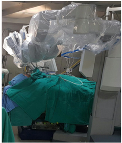
Figure 3: Docking of the robot for transoral surgery.

Lupinepublishers-openaccess-otolaryngology-journal

Conclusion: Excised swelling base of tongue - LINGUAL THYROID.

Results

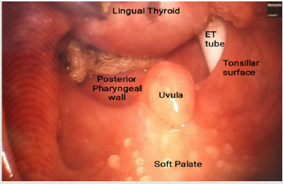
TORS of ectopic lingual thyroid tissue was successfully performed with excellent functional outcomes and with decreased operating time than conventional tongue base surgeries. Less than 5 ml blood loss during surgery and stay in the hospital was less than 24 hours (Figure 4).

Figure 4: Intraoperative picture of mass.

Lupinepublishers-openaccess-otolaryngology-journal

Discussion

Lingual thyroid is reported to be a rare congenital anomaly in the population. Asymptomatic ectopic thyroid tissue becomes symptomatic in periods of puberty, pregnancy, menopause, inflammation, and stress. Thyroid requirement increasing in these periods leads TSH level to increase and hypertrophy in the thyroid gland [8]. Approach to lingual thyroid tissue may vary according to clinical findings, but it is still debatable. Although asymptomatic cases can be followed without management for probable complications, in symptomatic cases, there are alternative treatment methods. Even though thyroid suppressive treatment conducted with the administration of exogenous thyroid hormone is a method that can be preferred in patients with minimal symptoms, it is not preferred in that it requires long term treatment. Radioactive iodine treatment and surgery are other treatment alternatives. As it is in a region difficult to access, open surgical approaches have been used in surgical treatment [9]. These surgical approaches may lead morbidity rates to increase according to the method used since they require lip, tongue, and mandibular splitting or lateral, suprahyoid pharyngotomy and long operation times and feeding with nasogastric tube, they run the risk of the development of suprahyoid pharyngotomy, hospitalization is prolonged, and there are skin scars. In order to avoid these morbidities, transoral resection techniques including Co2 laser, electrocautery, and harmonic technologies are used.
Terris et al. [10] reported that transoral minimal invasive lingual thyroid resection decreased morbidity and duration of hospitalization compared to other open approaches. Coblation assisted lingual thyroid and lingual tonsil resection techniques were used and successful results were reported [11-13]. Leitzbach et al. [12] reported successful results and low complication rates in 108 patients undergoing coblation assisted resection due to tongue root and lingual tonsil hypertrophy. However, transoral resection techniques have limited point of view and lead to difficulty in resection owing to difficulty in manipulation, which restricts their use [6]. Studies have demonstrated that as the size of the mass increases, transoral respectability of the mass decreases. The basic principles of avoiding complications in transoral tongue root resections are careful dissection in accordance with imaging methods and adequate anatomic evaluation in order to prevent lingual artery injury [14]. Transoral robotic surgery decreases the technical difficulties encountered in other transoral approaches in that it has a three-dimensional larger point of view, and manipulation is easier thanks to robotic instruments, leading to its larger use in oropharyngeal and tongue root operations [7]. Transoral robotic surgery is used in many different regions in surgical otolaryngology [15,16]. Three robotic arms, 0-degree and 30-degree high definition and 3-dimensional video instruments make it possible to overcome restriction of visualization of regions with difficult access and difficulty of manipulation. Lingual thyroid excision using this method has been described in previous studies [17,18]. The absolute local contraindication is related to the difficult exposition of the oropharynx, due to a limited opening of the mouth or the patient’s habitus. TORS for treatment of ectopic lingual goiter is feasible, requires a short learning curve, and appears to be safe [19]. In addition, the robotic surgery also has been adopted to do esthetic neck surgery with better cosmetic outcomes by hiding the scar [20].

Conclusion

TORS assisted excision of ectopic lingual thyroid is minimally invasive, feasible method. It is a safe technique with minimum morbidity and decreased operative time. In experienced hands it should be offered as valid treatment for this pathology. Studies with larger case series and longer duration of follow-up are required in order to reach a definitive conclusion on this issue.

Read More Lupine Publishers Otolaryngology Journal Articles:
https://lupine-publishers-otolaryngology.blogspot.com/
 

Saturday, June 12, 2021

Lupine Publishers | А Brief Algorithm of the Medel Cochlear Implant Fitting

 Lupine Publishers | Journal of Otolaryngology


Abstract

This article presents a brief algorithm of a cochlear implant fitting from the first fitting to the end of the first session of the implant setting. Most of the articles in the bibliography were written specifically for the future “Guidelines for cochlear implant setup”. Links to the world literature are in the references to the above-mentioned articles.

Procedure

a. Switch on the “Maestro” program and connect the processor to the DIB (Medel).

b. Fill the patient data, then place an antenna on the patient’s head, read the parameters of the implant [1].

c. Save the patient data and the implant parameters. Carry on telemetry and record its results in a speech processor.

d. Select the frequency range (250-6500 Hz) [2], set an activation level (95%), a coding strategy (FS4). The parameters of electrical stimuli should be set to the following: duration - 300ms, interval between stimuli - 600ms [1].

e. Proceed with sequential stimulation of all channels at small electrical levels in the SWEEP mode [1] with an observation of the patient’s response. A gradual increase of stimulation levels with three steps each, SWEEP stimulation and save the sequential programs. Step is 0.2–0.3dB.

f. If the patient’s response is detected, the first fitting will end. Afterwards create a map of three steps above the one where the response was detected. Create a configuration with these 4 maps. The fourth map is the last created map. 3, 2 and 1 maps are less in increments of 3 steps. We instruct to switch programs till the setting up the next day. We give our “Instruction” [3] in case of its absence.

g. In the following days, continue parallel increase of the levels of stimuli along all channels with SWEEP stimulation. Monitor the reaction of the patient and take into account the comments of parents and teachers. Remember the intraoperative levels of the stapedial reflex.

h. Train Categorical Loudness Scaling (CLS) in the categories “Hear” - “No sound” [4].

i. Relaxometry is performed when approaching the levels of the intraoperative reflex. N.B. Intraoperative - visual - threshold levels of stapedial reflex can be higher than real ones obtained on the impedance meter during the fitting, therefore it is impossible to fully rely on intraoperative threshold levels. We observe reactions of a child and take into account the comments of parents and teachers.

j. Register threshold reflex levels using the SWEEP stimulation-registration program and use them to set C-levels [5]. Based on the results of reflexometry and C-levels of the work program chosen by the parents, create a map which will be the second in the configuration and from that create one program lower by 3 steps (first map) and two program of increasing levels (3-rd and 4-th maps). The electrical threshold levels are set at 20 dB below the MCL-10% of the MCL.

k. Train CLS in the categories “No sound”, “Soft”, “Good”, “Loud” [4].

l. Parents and teachers choose a work program.

m. Using the SHCHUP program, determine the maximum comfortable sound pressure level of step noises [6]. The CLS experience helps the child. According to the results of the SHCHUP, adjust the current C-levels through the channels. After that configuration with four maps can be created - the second map is in accordance with the results of the SHCHUP, considering the C-levels of the comfort program chosen by the parents.

n. In order to choose a work program, we give an instructionexplanation to the parents.

“As you yourself understand, we all go on the same program. Sometimes we hear loudly. But we do not use ear plugs around the life. Why can’t an implanted patient hear loudly sometimes? Can. And he should. Sometimes! Therefore, the program at which the patient sometimes hear loudly is optimal- working one” [3].

o. During the session, use MIMIC program to demonstrate to the parents vootiue (on their own ear) the problems of CI patients and explain to them some features of the auditory perception of their children [7,8].

p. As the last step, carry out a telemetry, open the programs in turn, and agree to the possible proposal of “Maestro” to replace telemetry. Then write the new values of resistance of the electrodes in 4 programs. Afterwards record these programs with the latest telemetry, without changing the C-levels of the last configuration maps.

q. The first fitting session is completed.

Read More Lupine Publishers Otolaryngology Journal Articles:
https://lupine-publishers-otolaryngology.blogspot.com/


Choanal Atresia Repair, A Comparison Between Transnasal Puncture With Dilatation And Stentless Endoscopic Transnasal Drilling

  Abstract Background: in this study we present the outcome of surgical repair of choanal atresia of 33 patients underwent t...