Friday, December 25, 2020

Lupine Publishers | Evaluation of Ear, Nose and Throat Foreign Bodies in the Department of ENT-Head and Neck Surgery in a Teaching Hospital

 Lupine Publishers | Journal of Otolaryngology


Abstract

Objective: FB (foreign bodies) in ear, nose and throat are often encountered by otolaryngologists in their daily practice and is commonly seen in both children and adults. Depending upon the type and location of FB, it may have serious impact on individual’s health if instant appropriate action is not taken. That’s why, there’s frequent visits to ED (emergency department) on having FB in ear and aerodigestive tracts. The objective of this study was to evaluate the nature, common sites, modes of presentation, modes of management of FB, age and gender distribution.

Materials and Methods: A retrospective hospital-based study was done in Universal College of Medical Sciences, Bhairahawa, Rupandehi, Nepal from March 2014 to September 2017. The information was obtained from hospital record books.

Results: Out of 483 total patients, 287 (59.42%) were male and 196 (40.57%) were female. Most of them were less than 10 years old. Of the 483 patients, 202 (41.82%) had FB in the ear, 132 (27.32%) in the nose and 149 (30.84%) in the throat. Living FB were found in 54 (26.73%) patients out of 202 in the ear, 10 (7.57%) patients out of 132 in the nose and none in the throat. Of the total patients, 97 (20.08%) required general anesthesia (GA) to remove FBs and the rest 427 (88.4%) patients were dealt with or without local anesthesia. Most of the FBs were removed promptly on presentation otherwise within 24 hours of presentation in the hospital.

Conclusion: FB in ENT were found more commonly in the children and the commonest site was ear. Timely presentation, prompt diagnosis and needful management in a center with otolaryngology practice reduces the morbidity and mortality. Most of the FB in ENT can be removed in outpatient department (OPD) or emergency room (ER) with or without local anesthesia (LA).

Keywords: Ear; Nose; Throat; Foreign Bodies; Local Anesthesia

Introduction

A foreign body (FB) is any object or substance that is not derived from the individual’s own body part and can cause harm by its mere presence if prompt medical care is not provided [1,2]. They may be found in Ear, Nose and Throat. They are very common in otorhinolaryngological clinical practice. It can be introduced spontaneously or accidentally by both children and adults. However, children are common victims as they have habit of inserting nearby objects in their nose, ear or mouth, imitation and also other contributing factors are like boredom, playing, mental retardation, insanity, and attention deficit hyperactivity disorder, along with availability of the objects and absence of watchful caregivers. Consequently, it may cause minor irritation to life threatening problem. A proper technique, good light, appropriate instrument, a co-operative or fully restrained patient and a gentle approach by the related doctor or health worker are required for the removal of FB. One should have a clear diagnosis before making attempts to remove the FB so as to lessen the morbidity [3,4]. FB may be classified as animate (living) and inanimate (nonliving). The inanimate FB can again be classified as vegetative (organic) and non-vegetative (inorganic) FB, and hygroscopic (hydrophilic) and non-hygroscopic (hydrophobic) [1,2]. The objective of this study was to evaluate the nature, common sites, modes of presentation, modes of management of FB, age and gender distribution.

Materials and Methods

A retrospective study was conducted in the Department of ENT – Head and Neck Surgery, Universal College of Medical Sciences, from March 2014 to September 2017. The data were obtained from the hospital record books. Otoscopy and anterior rhinoscopy were performed to diagnose FB of the ear and nose respectively. Instruments such as Jobson Horne probe, FB hook, Tilley’s forceps, and crocodile forceps were used in FB removal from the nose and ear. Syringing and suctioning were also done for FB ear removal. Plain X-ray of the neck was done in patients with a history of FB ingestion. Flexible nasopharyngo laryngoscopy (NPL) and flexible upper gastrointestinal (UGI) endoscopy were done in cases where the FB was not visible in X-ray to rule out presence of a FB or to determine its site of impaction and in selected cases UGI endoscopy was used for FB removal too. It was followed by removal of the FB from the oropharynx/hypopharynx and esophagus with direct laryngoscopy or rigid esophagoscopy, respectively under general anesthesia (GA). FB struck in the oropharynx or parts of hypopharynx were confirmed with the help of Lack’s tongue depressor and head light or indirect laryngoscopy and removed with the Tilley’s forceps under local anesthesia (LA) in the OPD with patient co-operation.

Results

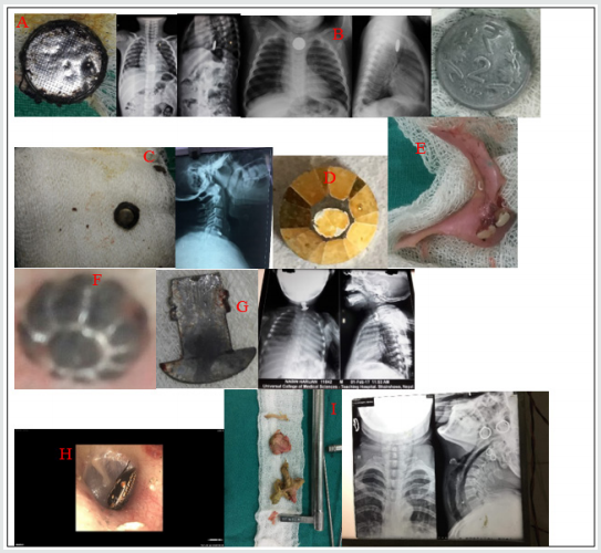
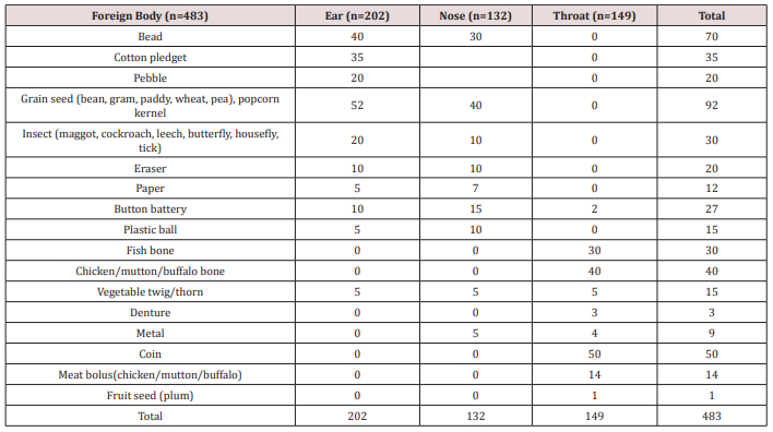
There were 483 patients recorded, out of which 287 (59.42%) were male and 196 (40.57%) were female with male to female ratio 1.46:1. The number of FB (Table 1 & Figure 1) in ear was 202 (41.82%), 132 (27.32%) in nose and 149 (30.82%) in the throat. Out of 202 FB in ear, 54 (26.73%) was animate and 148 (73.26%) inanimate. Out of 132 nasal FB, 10 (7.57%) was animate and 122 (92.42%) was inanimate. The FB encountered in throat was entirely of inanimate nature.

Figure 1. Few examples of ENT foreign bodies. A) FB toy battery in esophagus and removed with rigid esophagoscope, B) FB coin in esophagus, C) FB button battery in esophagus, D) FB bead removed from ear, E) FB denture, F) FB bead removed from nose, G) FB metal hook in esophagus, H) FB insect in ear and I) FB chicken bone with meat bolus in esophagus.
lupinepublishers-openaccess-journal-otolaryngology

FB in Ear

Total of 202 patients were recorded with FBs in the ear. The most common type of FB in the ear was grain seed 25.74% (52) followed by FB bead 19.8% (40). Fifty-four (26.73%) patients had animate (living) FBs. These were mostly insects in the form of maggots, cockroach, grasshopper, butterfly, housefly and ticks. Hundred and forty-eight (73.26%) patients had inanimate (nonliving) FBs. Out of 148 patients, fifty-two (35.13%) had hygroscopic FBs in the form grain seed in the form of bean, pea, wheat, paddy and gram as shown in Table 1. The rest ninety-six (64.86%) patients had non-hygroscopic FBs in the form of bead, cotton pledget, pebble, eraser, paper, button batteries, plastic ball and vegetable twig or thorn. Majority of cases were seen in 0-10 years age group i.e. 72% (147). The most common site of FB lodgement was found to be the external auditory canal. Most of these FBs were removed in the OPD or in the ER with or without local anesthesia (LA). In 4 children, the FB were found impacted in the deeper part of EAC (3 button batteries and 1 plastic ball) and had to remove under GA via post-aural approach.

FB in the Nose

Hundred and thirty-two (27.32%) had FB lodgement in the nose. The most common was grain seed 40 (Out of it, only 10 (7.57%) patients had living FBs i.e., 9 had maggots and 1 had leech. The rest 122 (92.42%) patients had nonliving FBs as shown in Table 1. Forty FB (30.3%) were of hygroscopic nature in the form of grain seed and the rest sixty nine percent being non-hygroscopic as in Table 1. Of the total number of 132 patients 122 patients (92.42%) were children and the rest 10 (7.57%) patients were adults with animate type of FBs. Hundred and five (86.06%) of the children presented with history of FB insertion nose by their caretakers, while in 17 (12.87%) children neither the patients nor the caretakers were certain of FB insertion. Unknowingly, they were treated as a case of sinusitis due to complaints of nasal blockage, headache and unilateral fetid discharge by the pediatricians and primary care physicians, which was later, referred to our center and revealed to be forgotten FB. Sometimes, even one has to depend on imaging like x-rays /CT scans to rule out the FBs where the patients are unable to recall the events. Otherwise, most of the times the typical history provides clue for clinching the diagnosis. Most of the FB were removed in the ER and OPD with or without LA and only 3 cases (2.27%) required removal under GA i.e. 2 cases of beads and 1 case of grain seed which on manipulation went posteriorly and also patient being uncooperative.

Foreign Bodies in the Throat

A total of 149 patients presented with the complaint of ingestion of FB.  The most common type of FB was coin of one rupee, 2, 5 and 10 rupees and the common site of the impaction was cricopharyngeal junction in all the 50 patients (33.55%). The sites of other types of FB impaction were oral cavity, oropharynx, hypopharynx, thoracic esophagus and lower gastro-esophageal sphincter region. All the ingested FB were inanimate, with 84 (56.37%) being organic and 65 (43.62%) being inorganic. Organic FBs were meat bolus and bone (fish, chicken, mutton, and buffalo meat) and one of plum seed. The inorganic FBs included button battery, thorn, denture, coin, and metallic objects as shown in Table 1. Age less than 10 years old were the most common group with FB coin. FBs fish bone and vegetable twig/thorn lodged in oral cavity and oropharynx were removed under LA. Out of 40 FB coin, 10 were dislodged spontaneously via gastrointestinal route, 5 FB meat bolus and 7 FB chicken bone were removed by flexible endoscopy and the rest of the FBs were removed under GA without  postoperative complications.

Table 1. Different types of Foreign bodies (FB) in ENT.

lupinepublishers-openaccess-journal-otolaryngology

Discussion

In our study, we’ve found higher incidence of FB in children less than 10 years old in 66.04% (319) patients. This is consistent with study by Iseh and Yahya [5], Ogunleye AOA and Sogebi R [6], Ahmad M, et al. [7]. This may be consequent of children’s exploring habit and lodging objects into the natural orifices of body, accidentally or intentionally. We found 59.42% of patients to be male and 40.5% to be female with male: female ratio 1.46:1. The male: female ratio was shown to be 1:1.05 by Gregogri et al. [8] whereas it was 1:1.26 in the study of Ogunleye AO et al. [9] and 1.35:1 by Agrawal S, Ranjit A study [10]. This suggests male are more susceptible than female to foreign body insertion in the orifices. In this study we observed ears were the most common site of lodgement of foreign bodies (41.82%) followed by throat (27.32%) and nose (27.32%). Parajuli R [11] and Shrestha I, et al. [4] also found in their study ears as the most common site for impaction of foreign bodies followed by throat and nose.  The most common foreign body in the ear and nose was the variety of grain seeds like bean, pea, paddy, wheat, gram, maize and foreign body coin was highest in throat. Removal methods, most commonly used for ear, nose and throat FBs were similar to those presented by Parajuli R [11], in order of preference the alligator forceps, Jobson Horne probe, foreign body hook, Tilley’s forceps and ear syringing. No patient required endoscopy or indirect laryngoscopy to remove oropharyngeal FB. The need for general anesthesia to remove FB varies in literature, with percentages varying from 8.6% to 30% [12]. There were no complications reported post FB removal.

Conclusion

FB in ENT are common in both pediatric and adult population. Comparatively the children are seen to be more vulnerable to have ENT FB lodgement.  Significant complications may arise if FB in ENT are not taken care of immediately and skillful removal is must. Thus, proper care and watch must be provided by care takers or the family members in order to prevent the encounter of such objects, especially in pediatric group.

Read More Lupine Publishers Otolaryngology Journal Articles:
https://lupine-publishers-otolaryngology.blogspot.com/


Tuesday, December 22, 2020

Friday, December 18, 2020

Lupine Publishers: Lupine Publishers | Review of Ten Years’ Experienc...

Lupine Publishers: Lupine Publishers | Review of Ten Years’ Experienc...:  Lupine Publishers | Journal of Otolaryngology Abstract Endoscopic skull base surg...

Lupine Publishers | A Cerebellopontine Angle Epidermoid Cyst Presenting as Trigeminal Neuralgia: A Case Report

 Lupine Publishers | Journal of Otolaryngology


Abstract

Trigeminal neuralgia is the one of the most painful condition known to mankind, so much so that it has described as suicide illness. The diagnosis of Trigeminal neuralgia is clinically plausible due to its specific characteristics. Differentiating Classical form secondary Trigeminal neuralgia requires further evaluation with either a CT or MRI. A correct diagnosis is crucial, as patients can then follow a generally acknowledged treatment modality of either Medication or surgery. The aim of this article is to present a case of Trigeminal Neuralgia secondary to Cerebellopontine angle Epidermoid cyst in an elderly female patient. She had to be maintained with medicines alone as she was skeptical of undergoing brain surgery. This report also portrays that secondary trigeminal neuralgia can be managed long term with carbamazepine without major adverse effects.

Keywords: Epidermoid Cyst; Cerebellopontine Angle; Trigeminal Neuralgia

Introduction

Trigeminal neuralgia is the one of the most painful condition known to mankind, so much so that it has described as suicide illness [1]. The diagnosis of trigeminal neuralgia is clinically plausible due to its specific characteristics. According to leading researcher of trigeminal neuralgia, Prof. Joanna Zakrzewska’s, it is one of the few chronic pain conditions in which sufferers can be rendered pain-free either with medications or surgery [2]. Therefore, correct diagnosis is crucial, as patients can then follow a generally acknowledged treatment modality. The aim of this article is to present a case of Trigeminal Neuralgia secondary to Cerebellopontine angle Epidermoid cyst in an elderly female patient, who has refused undergoing surgery and had to be maintained with medicines alone. This also portrays that secondary trigeminal neuralgia can be managed long term with carbamezapine without major adverse effects.

Case Report

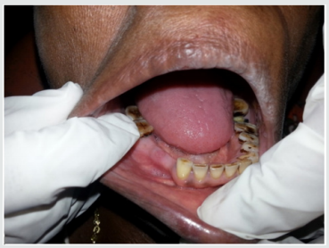
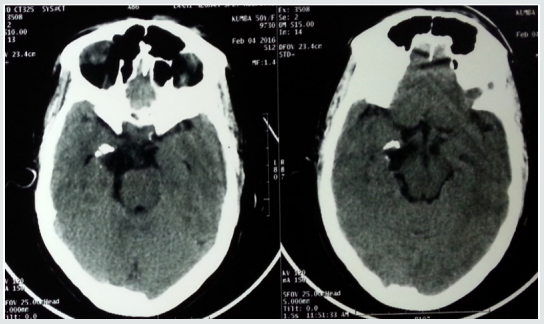
A 50-year-old female patient presented to the department of oral medicine and radiology, at Century dental college with complaint of excruciating pain in the lower right side of the face for one year. The pain was sudden in onset, sharp shooting in nature and lasted only for a few seconds. The episodes were frequent and occurred many times in a day. She had sought treatment at various dental clinic during which time most of her teeth on the right side were extracted (Figure 1). She had applied a skin oil for pain intraorally two days back which had resulted in chemical burn. Her medical history was significant, that she was under treatment for epilepsy and suffered with herpes zoster and was treated with acyclovir 800mg for 7 days. She was conscious and cooperative, of short stature and moderately nourished. There was pallor of the skin and inferior palpebra. Intraorally, pallor was noticeable on the buccal mucosa, erosion was detected on the lower right alveolar ridge region in relation to the missing premolars. The upper and lower arches were partially edentulous. Trigger zone was elicited at the right lower border of the mandible. A provisional diagnosis of Trigeminal neuralgia was made. CT scan of the brain showed a heterogenous lesion with cystic areas and calcification in right Cerbello Pontine angle extending to the medial aspect of temporal lobe. There is compression and rotation of brain stem to the left. An impression of CP angle neoplasm was given on CT (Figure 2).

Figure 1.

lupinepublishers-openaccess-journal-otolaryngology

Figure 2.

lupinepublishers-openaccess-journal-otolaryngology

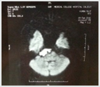
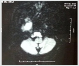
MRI of the brain revealed a lesion of size 15X33X30mm involving the right CP angle cistern, which appeared hypointense in T1 (Figure 3a) and hyperintense in T2 image. It was causing compression of pons and lower midbrain. The 3rd and 6th cranial nerves were compressed and encased by the lesion with displacement towards left. 7th and 8th cranial nerves were closely abutted (Figure 3b). MR study was suggestive of epidermoid cyst involving right basal cistern and CP angle cistern. Based on these findings, diagnosis of Trigeminal neuralgia secondary to epidermoid cyst located at right cerebella pontine cistern was made. Patient was prescribed carbamezapine 200mg twice daily gradually increased to 400mg tid. She was referred to neurologist who advised surgery for the brain lesion. Patient and her family members were very apprehensive about surgery and did not want to proceed with surgery. She has been on medication for the past 2 years and the dose of carbamezapine was reduced to 100mg twice daily. Regular blood test has been done to check for myelosuppression. There was a single episode of unbearable pain, for which inferior alveolar nerve block was given to arrest the pain. Long term treatment follows up of the patient can be described as favorable as she has been able to carry on normal routine with only few occasional episodes of severe pain.

Figure 3a: Shows a transverse fracture of petrous bone crossing the turns of vestibule.

lupinepublishers-openaccess-journal-otolaryngology

Figure 3b: Another case of transverse fracture. A Transverse fracture of petrous bone is seen reaching horizontal semi-circular canal and causing hemotympanum.

lupinepublishers-openaccess-journal-otolaryngology

Discussion

Trigeminal neuralgia is a neuropathic pain with distinct diagnostic criteria. The IASP definition of trigeminal neuralgia (TN) is “sudden, usually unilateral, severe, brief, stabbing, recurrent episodes of pain in the distribution of one or more branches of the trigeminal nerve.” Trigeminal neuralgia is a rare condition [2]. The peak incidence is in the age group of 50 – 60 with preponderance in women [3]. However, some Japanese and Chinese reports have quoted a male predominance in those above 80 years of age [4]. It is a condition that has been recognized many centuries ago, The French terminology Tic douloureux was coined by surgeon Nicolaus Andre [5]. There are two recognized forms of TgN, Classical and Secondary or Symptomatic which is related either to central nervous system lesion or multiple sclerosis [4]. There is also Atypical form which does not have clearly definable features and symptoms may overlap that of atypical facial pain. This has been described as atypical trigeminal neuralgia4. The aetiology of the condition is still unclear5. Most researchers suggest that root entry zone (REZ) of the trigeminal ganglion to be site of generation of pain. Compression of the REZ by blood vessels or tumors and demyelination are the most common abnormality found in this area. This leads to hyperexcitability and central sensitization [2,4,5].

Trigeminal neuralgia is a clinical diagnosis. The characteristic features being

a) Paroxysmal (abrupt onset and termination) attacks of pain lasting for a few seconds to few minutes

b) It involves one or more division of trigeminal nerve, unilaterally.

c) Character is described as sharp, shooting, stabbing or burning.

d) Severe intensity.

e) Presence of Trigger zones (stimulus evoked: palpation by routine activity such as touching, washing the face, brushing teeth) intensifies pain usually found around the nose or mouth.

f) Presence of refractory period.

The two accepted modalities for treatment are medical and surgical.

Medical Management

Gold standard for medical treatment is response of the condition to Carbamazepine. Carbamazepine is a tricyclic imipramine first synthesized in 1961 and introduced for treatment of trigeminal neuralgia by Blom [5,6]. It is usually started at 200mg per day single dosing and gradually increased to up to 800mg i.e. four divided doses of 200mg each. The most common side effect is hypersensitivity (5 – 10 % of the patient), folic acid deficiency and megaloblastic anemia6. Drug interaction with warfarin has also been reported. Oxycarbazepine, is a daughter drug and has fewer side effects. A 300mg is equipotent to 200mg of carbamazepine. The improved safety profile makes it a better option to use. Lamotrigine, Gabapentin and Baclofen are other second-line drugs that could be prescribed [2,5,7].

Surgical Procedures

Various surgical procedures have been conducted and they can be briefly classified into

a) Peripheral surgery such as Neurectomies, Cryotherapy, Laser, Radiofrequency Thermo-Rhizotomy and Injections with Streptomycin, Alcohol, Glycerol and Phenol. Analgesic blocking of peripheral receptors also arrests pain. This effect is also diagnostic [8].

b) Surgery at Gasserion Ganglion: Percutaneous radiofrequency rhizotomy /thermocoagulation and Percutaneous glycerol rhizolysis. These procedures are less invasive and are associated with low rates of mortality and morbidity. However, they are associated with anesthesia dolorosa, facial numbness and dysesthesia [8].

c) Surgery at the REZ: Microvascular decompression is one of the most successful procedures, providing relief for 70% of patients for up to 10 years. It is a nondestructive procedure. This procedure involves major neurosurgery and is therefore not suitable for all patients; it carries a mortality rate of 0.5% and a 2% risk of hearing loss [9]. Gamma knife surgery is a noninvasive radiosurgery [8].

All forms of surgery have potential for complications with sensory loss being the most common one. Recurrence of pain within 1- 4 years occurs with most of the procedures. The reported range for TN due to tumors is 0.8%-11.6% [10]. Tumors leading to TN are mostly benign and typically compress the root near its entry into the pons. Epidermoid tumors are slow growing, and symptoms also appear later in the course, it is also referred to as Cholesteatoma [11]. Patients usually present in 4- 5th decade of life Hearing loss is the most common presenting sign followed by trigeminal neuralgia. Other neurological deficits include facial paresis, hearing disturbances and third and sixth nerve palsy. It has been suggested that if pain occurs bilaterally or there is simultaneous involvement with other nerve trunk then a systemic involvement or expanding tumor has to be suspected [12]. Pathogenesis of TgN in epidermoid is uncertain, however it has been attributed to either direct compression or displacement of the nerve at REZ. It has been reported that neuralgia due to epidermoid tumors are clinically indistinguishable to classical TgN. However, the age of onset in this condition is earlier than the Classical TgN [12]. In this case the symptoms started at the age of 49 years and were confined to mandibular branch of trigeminal nerve only, it remained rather consistent with only sporadic flareup. This may be attributed to the medical treatment (carbamzepine).

On CT scans they are homogenous density of an epidermoid cyst enables it to be distinguished from other tumors [12]. In MRI they have lamellated or onion skin appearance. They have low or intermediate intensity in T 1 weighted images and high intensity in T2 weighted images13. MRI is the preferred imaging modality to visualize the anatomic landmarks around the trigeminal ganglion and the CP angle as it gives the best soft-tissue resolution and excellent visualization of the intracranial and extracranial course of the nerve [12,13]. They have to be differentiated from Schwannomas, meningiomas or chondromas by signal intensity criteria [13]. Surgically the cyst appears as white and pearly with encasement or compression of the trigeminal nerve. Optimum treatment is radical removal of the complete tumor [14]. This is problematic in most cases as it has wide extension and firm adhesion to neurovascular structures. To minimize neurologic deficits a part of the tumor is left behind. Other complications that are reported are meningitis, cerebellar and brain stem infarction. The possible REZ arterial compression is treated with an additional Microvascular decompression. 0 -30% estimated recurrence rate have been reported in long follow up studies [14]. One of the main aspects regarding trigeminal neuralgia as highlighted by Drangsholt and Truelove is the diagnosis of this condition [15]. Majority of patients attending the clinic have previously been misdiagnosed and had undergone irreversible dental treatment, even when they had presented with classical features of TN, as in this case.

Conclusion

William Osler once said, “Listen to the patient: he is telling you the diagnosis.” There are no objective diagnostic tests for idiopathic TNA. As a part of the diagnostic work-up, patients should have a magnetic resonance imaging (MRI) scan or a computed tomography scan to rule out secondary TN. If detected, these lesions are best treated surgically. although surgical procedures do carry risk of significant morbidity. However, convincing the patients regarding surgical treatment may be difficult as in this case report. Patients should be treated with carbamezapine as a first line therapy, followed by addition of other agents if patients become unresponsive. The patients must be followed up for long term for any adverse effects.

Funding

This research did not receive any specific grant from funding agencies in the public, commercial or not for profit sectors.

Read More Lupine Publishers Otolaryngology Journal Articles:
https://lupine-publishers-otolaryngology.blogspot.com/


Choanal Atresia Repair, A Comparison Between Transnasal Puncture With Dilatation And Stentless Endoscopic Transnasal Drilling

  Abstract Background: in this study we present the outcome of surgical repair of choanal atresia of 33 patients underwent t...