Monday, March 16, 2020

Lupine Publishers | Statistical Software: A Risk for Medical Science?

Lupine Publishers | Journal of Otolaryngology Research Impact Factor


Opinion

pinion This editorial is based on my personal professional experiences of some four decades work in health and business applications of statistics. I am fully aware that these experiences cannot be interpreted as a random sample and there is no warranty of any kind for the absence of possible biases. You might well be familiar with the US-FDA view that there is no unbiased data in the scientific universe available. Historically, the use of statistics in medicine is a well-documented fact since about two centuries. My personal experiences cover the transition period from electronic desktop calculators with paper and pencil, to the omnipresence of cheap computing power and sophisticated statistical software of today. I remember my early professional study design activities as a statistician as heavily impacted by cost considerations: Human work time was and still is quite expensive. During the last period of about some two decades I got the strong impression that the medical profession, especially those doctors who are working scientifically – showed a quite strong trend to higher statistical understanding and knowledge as compared to my early working times. This positive trend is highly appreciated by me and I see it as an enormous economic advantage that computers took over the tedious workload of numbers crunching nowadays. There is a negative trend involved over the last two decades as well: I observed a growing trend of numbers of “so called” statisticians who actually are experts in using available statistical software packages only but have little or no statistical expertise. I see a professional statistician as a human who understands the primary objective of the study’s objective and to assess the medical question under consideration and to decide the statistical model selection for the very question based on the scope of the scientific question and all of the constraints and limitations in practice. I know that this is an “ideal world” assumption and we all are but humans with our limitations. My experience that it pays to strive for perfection is illustrated by some examples which I consider to be potentially useful and beneficial for your work as doctor:

Project Planning Phase

a) Overall considerations of project management and quality control, legal requirements.
b) Definition of research question(s).
c) Definition of data selection criteria, sampling variables and observation time(s) schedule.
d) Aspects of data collection and documentation, ongoing project quality controls.
e) Feasibility considerations of various project designs.
f) Administrative aspects: financials, selection of partners, estimation of required realization times.
g) Statistical methods for data analysis, feasibility of pilot- (sub)-projects.
h) Final definition of data selection criteria.
i) Aspects of results publication.
j) Aspects of possibly necessary actions in case of emergencies.

Project Realization Phase

a) Ongoing control of “plan vs actual” progress.
b) Ongoing communication between all project partners.
c) Maintenance of open minds for early signs of project’s critical developments.
d) Ongoing monitoring for possible emergency actions.
e) The KISS principle: Keep everything as simple and stupid as possible.
I’d like to recommend using the items in the above project management as guidance for your project but under no condition as a comprehensive cookbook! As ENT scientist you should permanently remember that you are doing research in humans and not in bolts and nuts!

How you could easily detect the difference between a
“user of statistical software” or a “real” statistician

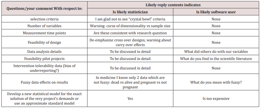
                         The next table provides you with selected questions/topics to distinguish between professional statistician and software user (Table 1).
Table 1.lupinepublishers-openaccess-journal-otolaryngology


Tuesday, March 3, 2020

Monday, March 2, 2020

Lupine Publishers | Post Endodontic Pain Reduction using three Irrigants with Different Temperature

Lupine Publishers | Journal of Otolaryngology Research Impact Factor

Abstract

Objective: The purpose of this research was to evaluate whether meticulous irrigation with three different temperatures would help in a decrease dental pain.
Materials and Methods: All 120 patients had teeth chosen for conventional RCT for prosthetic reasons in teeth with vital pulps. All canals were cleaned and shaped with Reciprocal files. Final irrigation was done with cold saline solution (6 OC, 4 OC, and room temperature).
Results: A total of 120 of 135 patients (69 females and 51 male) were included whereas 15 were excluded as not achieving the necessities of the study. All patients presented with a vital upper or lower molar, premolar, or front teeth. No statistically major change (P>0.05) between the groups was found regarding the degree or duration of pain.
Conclusion: The approach in both selecting the patients participating in the research and analyzing the data in this research allows us to determine that cryotherapy is an aid of clinical procedures to clean and shape the canals to decrease the occurrence of post-endodontic pain and the need for medication in patients presenting with a diagnosis of vital pulp.
Keywords: Apical healing; Flare-ups; Pain; Post endodontic pain; Post-operative pain

Introduction

Post-endodontic pain is an undesirable sensation occurred in patients regardless of the preoperative periapical status of the tooth treated. Therefore, prevention and management of post endodontic pain are essential in endodontic practice [1]. Organic material, microorganisms, and irrigating solutions extruding beyond the apical constriction during root canal therapy (RCT) will originate inflammation and periodontal ligament complications, such as severe pain or flare-ups. It must be noticed that the amount of extruded material (debris and/or irrigate) varies widely in the reported studies which indicate problems and inconsistencies in treatment methodologies [2-4]. Recent literature has showed that keeping apical patency would not generate more postoperative difficulties [5-7]. A recently issued in vitro study showed that intracanal delivery of cold irrigating solution at 2.5 °C with negative pressure flushing reduced the external surface temperature to close 10 °C [8-10], would be enough to create a local anti-inflammatory beneficial consequence in peri radicular tissues. Cryotherapy proposes that using cold over some procedures may decrease the diffusion of nerve signs, bleeding, edema, and local inflammation and is therefore effective in the reducing of pain. Therefore, the purpose of this research was to evaluate whether meticulous irrigation with three irrigating practices with different temperature would help in a decrease of post-endodontic pain.
Three expert endodontists with a private practice of 17 years and skilled in the procedures and procedures studied were included in the research and performed 40 RCTs each (a total of 120) in upper/lower front or back teeth with irreversible pulpitis recognized by pulp sensitivity testing with hot and cold.  Pulpal response tests were achieved by the main author, and a digital X ray diagnosis was documented by three certified clinicians. Additional clinical necessities for patients´ inclusion were as follows: Necessities of the research were agreed and spontaneously accepted, healthy patients were included, teeth with enough coronal structure and diagnosed with vital pulps, no previous RCT, and no analgesics or antibiotic consumption 7 days before the procedures. A total of 120 of 135 patients (69 females and 51 male) aged 18 – 60 years were referred and integrated in this research, whereas 15 were rejected as not accomplishing the necessities wanted. All participants showed with a vital upper or lower molar, premolar or front teeth designated for conventional RCT for dental rehabilitation reasons.

Methods

Dental procedures

Root canal treatment was done in one visit. Topical anesthetic (Anesthesia Topical, Astra, Mexico) was used. Patients received 2 carpules of articaine 2% with epinephrine 1:200,000 (Septodont, Saint-Maur des-Fosses, France). Situations in which supplementary anesthesia was needed, intra-ligamental anesthesia (2mL articaine 2%) was supplied. For the upper front teeth, the solution was administered by tender and slow local infiltration. For the lower teeth, one of the carpules was used for the lingual and alveolar nerve block, the other one for a moderate bucal infiltration nearby the tooth to be treated.

Irrigation protocols

Group 6 °C. The R25 (size 25/ .08) instrument was employed in tinny and curved canals, and R40 files (40/ .06) were used in broad root canals. Three in-and-out pecking series were employed with a fullness of not more than 3mm until getting the calculated WL. Patients allocated to this group receive a final irrigation with 5mL of cold (6 °C) 17% EDTA followed by 10mL of cold (6 °C) sterile saline solution dispensed to the WL using a cold (6 °C) metallic micro-cannula.
Group 4: Canals were instrumented as in group A. Patients allocated to this set received a final irrigation with 5mL of cold (4 °C) 17% EDTA followed by 10mL of cold (4°C) sterile saline solution  dispensed to the WL using a cold (4 °C) metallic micro-cannula for 1 minute.
Group RT: The R25 (size 25/ .08) instrument was employed in tinny and curved root canals, and R40 files (40/ .06) were used in wide canals. Three in-and-out series were employed with a space of not more than 3mm until getting the calculated WL. Reciprocal instruments were used in one tooth only (single use). Participants allocated to this control group were treated similarly to the experimental groups, except that they received a final flush with 5mL (room temperature) of 17% EDTA followed by 10 mL (room temperature) of sterile saline solution delivered to the WL.

Statistical analysis

The related issues preoperatively recorded were integrated into the examination as follows: age and sex, occlusal contacts, and maxilla or mandibular teeth. Changes in the strength of pain among groups were studied using the ordinal (linear) X2 test. Variances in VAS-recorded standards after 24, 48, and 72 hours and in the quantity of analgesic intake among the two groups tested.

Results

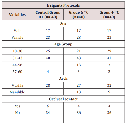
Table 1: Distribution by group of teeth and location.
lupinepublishers-openaccess-journal-otolaryngology
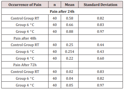
Table 2: Kruskal/Wallis test applied to the post-endodontic pain.
lupinepublishers-openaccess-journal-otolaryngology
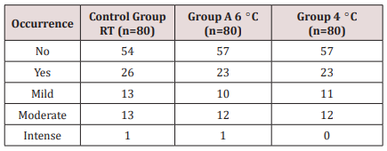
Table 1 displays the distribution of variables; a total of 120 participants took part in this study: 69 (57.5%) were women, and 51 (42.5%) were men. The ages fluctuated among 18 and 60 years; 87 (72.5%) were upper teeth, and 33 (27.5%) were lower teeth. The clinical management of the patients is showed in Table 1. No significant modification (P > 0.05) between the groups was encountered concerning the grade or period of pain. Rendering to the VAS examination, marks were seen 24 – 72 hours late in the 3 groups with a significant decline successively (Tables 2 & 3).
Table 3: Kruskal/Wallis test applied to the post-endodontic pain.
lupinepublishers-openaccess-journal-otolaryngology

Discussion

Pain is tough to comprehend and calculate especially when it occurs unexpectedly in patients. The major trouble in knowledge painful and discomfort is the participant’s individual valuation and its dimension. For this objective, organization of the estimation form has to be entirely understood by participants. In our research, a simple spoken classification was followed in the feedback procedure with four classes: no pain, slight, modest, and intense pain. These classes were clearly comprehended by participants and were described by the occurrence or nonappearance of the necessity for pain-relieving treatment. Preoperative pain is one of the main predictors of post-endodontic pain [11-14]. Thus, only teeth with irreversible pulpitis indicated for RCT because of prosthodontic purposes were treated in this research. In our research, we reduced the variation in the procedures following protocols based on recommendations by authors and manufacturers. While successful endodontic treatment depends on various variables, an important point to consider in the shaping of the root canal system is the amount of the irrigating solution. Proper disinfecting and filling the root canal system is facilitated by the keeping of its original shape from the entrance to the apical third, without any iatrogenic event.

Conclusion

According to the conditions established for this study, there was no statistically significant difference between the instrumentation systems assessed.

Lupine Publishers: Acute Liver Failure and Thyrotoxicosis Managed wit...

Lupine Publishers: Acute Liver Failure and Thyrotoxicosis Managed wit...: Journal of Surgery | Lupine Publishers Abstract Acute liver failure and hyperthyroidism are not typically common, although ...

Choanal Atresia Repair, A Comparison Between Transnasal Puncture With Dilatation And Stentless Endoscopic Transnasal Drilling

  Abstract Background: in this study we present the outcome of surgical repair of choanal atresia of 33 patients underwent t...